Medical Ultrasound Imaging
Sunday, 28 April 2024
Info Sheets Out- side     | 'Cine' Searchterm 'Cine' found in 44 articles 3 terms [ • ] - 41 definitions [• ] Result Pages : •
A cineloop is a period of images, stored digitally as a sequence of individual frames. Cineloops recorded at high frame rates may contain more frames than were displayed during the examination.
•  European Medicines Agency (EMA) is a decentralized body of the European Union with headquarters in Amsterdam.
'The EMEA began its activities in 1995, when the European system for authorizing medicinal products was introduced, providing for a centralized and a mutual recognition procedure. The EMEA has a role in both, but is primarily involved in the centralized procedure. Where the centralized procedure is used, companies submit one single marketing authorization application to the EMEA. A single evaluation is carried out through the Committee for Medicinal Products for Human Use (CHMP) or Committee for Medicinal Products for Veterinary Use (CVMP). If the relevant Committee concludes that quality, safety and efficacy of the medicinal product is sufficiently proven, it adopts a positive opinion. This is sent to the Commission to be transformed into a single market authorization valid for the whole of the European Union.' 'Its main responsibility is the protection and promotion of public and animal health, through the evaluation and supervision of medicines for human and veterinary use. The EMEA coordinates the evaluation and supervision of medicinal products throughout the European Union. The Agency brings together the scientific resources of the 25 EU Member States in a network of 42 national competent authorities. It cooperates closely with international partners, reinforcing the EU contribution to global harmonization.' See also Class I II III Devices, Phase 1 2 3 4 Drug Trials, and Drug Development and Approval Process USA.
Contact Information
MAIL
European Medicines Agency
Domenico Scarlattilaan 6 1083 HS Amsterdam The Netherlands
PHONE
+31 (0)88 781 6000
ONLINE
•
(DICOM) DICOM is the industry standard for transferral of radiologic images and other medical information between computers. Patterned after the Open System Interconnection of the International Standards Organization, DICOM enables digital communication between diagnostic and therapeutic equipment and systems from various manufacturers. The DICOM 3.0 standard evolved from versions 1.0 (1985) and 2.0 (1988) of a standard developed by the American College of Radiology (ACR) and National Electrical Manufacturers Association (NEMA). To support the implementation and demonstration of DICOM 3.0, the RSNA Electronic Communications Committee began to work with the ACR-NEMA MedPacs ad hoc section in 1992. Also Picture Archiving and Communication Systems (PACS), which are connected with the Radiology Information System (RIS), use commonly the DICOM standard for the transfer and storage of medical images. See also Digitization. Further Reading: Basics:
•
The definition of imaging is the visual representation of an object. Medical imaging is a broad term that encompasses various imaging modalities and techniques used in the field of medicine to visualize and study the body's anatomy and physiology. It includes both diagnostic and non-diagnostic imaging procedures, where diagnostic imaging specifically refers to the subset of medical imaging techniques that are primarily focused on diagnosing diseases or conditions. Medical imaging techniques are employed to obtain images or visual representations of the internal organs, tissues, and structures, aiding in the diagnosis, treatment, and monitoring of medical conditions.
The field of medical imaging has significantly evolved since the discovery of X-rays by Konrad Roentgen in 1896. Initially, radiological imaging involved focusing X-rays on the body and capturing the images on a single piece of film within a specialized cassette. Subsequent advancements introduced the use of fluorescent screens and special glasses for real-time visualization of X-ray images. A significant breakthrough came with the application of contrast agents, enhancing image contrast and improving organ visualization. In the 1950s, nuclear medicine studies utilizing gamma cameras demonstrated the uptake of low-level radioactive chemicals in organs, enabling the observation of biological processes in vivo. Currently, positron emission tomography (PET) and single photon emission computed tomography (SPECT) technologies play pivotal roles in clinical research and the diagnosis of biochemical and physiological processes. Additionally, the advent of the x-ray image intensifier in 1955 facilitated the capture and display of x-ray movies. In the 1960s, diagnostic imaging incorporated the principles of sonar, using ultrasonic waves generated by a quartz crystal. These waves, reflecting at the interfaces between different tissues, were received by ultrasound machines and translated into images through computer algorithms and reconstruction software. Ultrasound (ultrasonography) has become an indispensable diagnostic tool across various medical specialties, with immense potential for further advancements such as targeted contrast imaging, real-time 3D or 4D ultrasound, and molecular imaging. The first use of ultrasound contrast agents (USCA) dates back to 1968. Digital imaging techniques were introduced in the 1970s, revolutionizing conventional fluoroscopic image intensifiers. Godfrey Hounsfield's pioneering work led to the development of the first computed tomography (CT) scanner. Digital images are now electronic snapshots represented as grids of dots or pixels. X-ray CT brought about a breakthrough in medical imaging by providing cross-sectional images of the human body with high contrast between different types of soft tissue. These advancements were made possible by analog-to-digital converters and computers. The introduction of multislice spiral CT technology dramatically expanded the clinical applications of CT scans. The first magnetic resonance imaging (MRI) devices were tested on clinical patients in 1980. With technological improvements, such as higher field strength, more open MRI magnets, faster gradient systems, and novel data-acquisition techniques, MRI has emerged as a real-time interactive imaging modality capable of providing detailed structural and functional information of the body. Today, imaging in medicine offers a wide range of modalities, including:
•
X-ray projection imaging;
•
Fluoroscopy;
•
Computed tomography (CT / CAT);
•
Ultrasound imaging (US)
•
Magnetic resonance imaging (MRI), Magnetic source imaging (MSI);
•
Single photon emission computed tomography (SPECT);
•
Positron emission tomography (PET);
•
Mammography.
These imaging modalities have become integral components of modern healthcare. With the rapid advancement of digital imaging, efficient management has become important, leading to the expansion of radiology information systems (RIS) and the adoption of Picture Archiving and Communication Systems (PACS) for digital image archiving. In telemedicine, real-time transmission of all medical image modalities from MRI to X-ray, CT and ultrasound has become the standard. The field of medical imaging continues to evolve, promising further innovations and advancements in the future, ultimately contributing to improved patient care and diagnostics. See also History of Ultrasound Contrast Agents, and History of Ultrasound. Further Reading: News & More:
•  From Siemens Medical Systems;
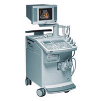
'This extremely flexible system supports targeted applications for OB/GYN, Radiology, Internal Medicine, Urology, and Prostate Brachytherapy. The large transducer selection, most with high-frequency capabilities, provides the right tool for general imaging, radiology, and internal medicine applications.'
Device Information and Specification
CLINICAL APPLICATION
OB/GYN, cerebrovascular, orthopedics, urology, peripheral vascular, pediatrics, small parts, surgery, abdominal, musculoskeletal
CONFIGURATION
Mobile, compact
DISPLAY MODES
Broadband, high-fidelity, multi-frequency, linear and curved
PROBE PORTS
Two - four
256
MEASUREMENT/CALCULATION FUNCTIONS
OB/GYN measurements and report package, off-line analysis
OPTIONAL PACKAGE
IMAGE PROCESSING
Result Pages : | Share This Page Look Ups |
Medical-Ultrasound-Imaging.com
former US-TIP.com
Member of SoftWays' Medical Imaging Group - MR-TIP • Radiology TIP • Medical-Ultrasound-Imaging
Copyright © 2008 - 2024 SoftWays. All rights reserved.
Terms of Use | Privacy Policy | Advertise With Us
former US-TIP.com
Member of SoftWays' Medical Imaging Group - MR-TIP • Radiology TIP • Medical-Ultrasound-Imaging
Copyright © 2008 - 2024 SoftWays. All rights reserved.
Terms of Use | Privacy Policy | Advertise With Us
[last update: 2023-11-06 01:42:00]