Medical Ultrasound Imaging
Sunday, 19 May 2024
Info Sheets Out- side     | 'Harmonic Power Doppler' p4 Searchterm 'Harmonic Power Doppler' found in 20 articles 1 term [ • ] - 4 definitions [• ] - 15 booleans [• ]Result Pages : •
Selective detection of the microbubble contrast medium can be enhanced by Doppler processing that removes signals with zero Doppler frequency shifts. This will remove tissue harmonics.
By detecting overlong bursts of inverted pulses and using Doppler detection methods, very high sensitivity to microbubbles can be achieved. The bubbles can be detected at sufficiently low incident power levels to avoid destroying them. Pulse inversion Doppler has demonstrated the first real-time images of myocardial perfusion using perfluorocarbon gas agents. See also Pulse Inversion Imaging, Myocardial Contrast Echocardiography, and Perfluorochemicals. Further Reading: Basics:
•
The thermal effect of ultrasound is caused by absorption of the ultrasound beam energy. As the ultrasound waves are absorbed, their energy is converted into heat. The higher the frequency, the greater the absorbed dose, converted to heat according the equation: f = 1/T where T is the period as in simple harmonic motion. Ultrasound is a mechanical energy in which a pressure wave travels through tissue. Heat is produced at the transducer surface and also tissue in the depth can be heated as ultrasound is absorbed. The thermal effect is highest in tissue with a high absorption coefficient, particularly in bone, and is low where there is little absorption. The temperature rise is also dependent on the thermal characteristics of the tissue (conduction of heat and perfusion), the ultrasound intensity and the length of examination time. The intensity is also dependent on the power output and the position of the tissue in the beam profile. The intensity at a particular point can be changed by many of the operator controls, for example power output, mode (B-mode, color flow, spectral Doppler), scan depth, focus, zoom and area of color flow imaging. The transducer face and tissue in contact with the transducer can be heated. See also Thermal Units Per Hour and Ultrasound Radiation Force. •
Ultrasound at the microbubble resonance frequency can cause bubble rupture at high acoustic power (mechanical index (MI) greater than 0.5). The result is a transient high-amplitude, broadband signal containing all frequencies, not only the harmonics. It will create a strong signal in B-mode or a short-lasting multicolored, mosaic-like effect in color Doppler sonography. Several terms for this typical signal have been used, e.g. induced or stimulated acoustic emission, loss of correlation imaging and sono-scintigraphy. •
[This entry is marked for removal.] From POINT Biomedical Corp CARDIOsphere® is an ultrasound contrast agent for assessment of myocardial perfusion in patients with coronary artery disease composed of highly echogenic bispheres. PB127 is a new developed microbubble with a bilayer polymer/albumin shell filled with nitrogen gas that has ideal characteristics for power harmonic Doppler. They can be destroyed by high power ultrasound, and spectral decorrelation between ultrasound pulses is maximized by rapid dissolution of the released nitrogen gas. POINT Biomedical Corp. announced (March 01, 2004) that it has completed two Phase 3 trials of CARDIOsphere®. The Phase 3 trials were designed to evaluate the performance of CARDIOsphere® imaging relative to radionuclide imaging for detecting obstructive coronary artery disease and identifying the anatomic location of perfusion defects.
Drug Information and Specification
RESEARCH NAME
PB 127
DEVELOPER
INDICATION -
DEVELOPMENT STAGE Myocardial perfusion -
Phase 3 completed APPLICATION
Infusion
TYPE
Microbubble
Polylactide/Albumin
CHARGE
Slight Negative
Nitrogen
PREPARATION
Reconstitute with 2ml H2O per vial and dilute with 150 ml DSW
DO NOT RELY ON THE INFORMATION PROVIDED HERE, THEY ARE
NOT A SUBSTITUTE FOR THE ACCOMPANYING PACKAGE INSERT! • 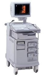 From ALOKA Co., Ltd.;
'The ProSound SSD-4000 utilizes the most advanced acoustic technologies available today, and its multidisciplinary technology architecture enables it to offer great versatility and flexibility over a wide range of clinical applications. With its new-generation, front-end technology including a 12-bit A/D converter, the ProSound SSD-4000 offers superior contrast resolution−especially when compared to 10-bit systems.'
Device Information and Specification
APPLICATIONS
CONFIGURATION
Compact, portable, dual dynamic display
Wide-band super high-density (W-SHD) transducers
OPTIONAL PACKAGE
Volume Mode
STORAGE, CONNECTIVITY, OS
Data Management Subsystem (iDMS), DICOM-Worklist
DATA PROCESSING
Result Pages : | Share This Page Look Ups |
Medical-Ultrasound-Imaging.com
former US-TIP.com
Member of SoftWays' Medical Imaging Group - MR-TIP • Radiology TIP • Medical-Ultrasound-Imaging
Copyright © 2008 - 2024 SoftWays. All rights reserved.
Terms of Use | Privacy Policy | Advertise With Us
former US-TIP.com
Member of SoftWays' Medical Imaging Group - MR-TIP • Radiology TIP • Medical-Ultrasound-Imaging
Copyright © 2008 - 2024 SoftWays. All rights reserved.
Terms of Use | Privacy Policy | Advertise With Us
[last update: 2023-11-06 01:42:00]