Medical Ultrasound Imaging
Sunday, 19 May 2024
Info Sheets Out- side     | 'Kidney Ultrasound' p3 Searchterm 'Kidney Ultrasound' found in 15 articles 1 term [ • ] - 3 definitions [• ] - 11 booleans [• ]Result Pages : •
(AUS) Abdominal ultrasound, also known as abdominal sonography, is a medical imaging technique that focuses on the visualization and assessment of the abdominal organs. While 'abdominal ultrasound' is the commonly used term, there are alternative terms that can be used to refer to this imaging modality: (TAE) transabdominal echography, abdominal ultrasonography, sonogram, FAST (Focused Assessment with Sonography for Trauma). Abdominal ultrasound imaging is an invaluable clinical tool for identifying the underlying cause of abdominal pain. An abdominal ultrasound examination encompasses a comprehensive evaluation of the liver, gallbladder, biliary tree, pancreas, spleen, kidneys, and abdominal blood vessels. It is a cost-effective, safe, and non-invasive medical imaging modality that is typically utilized as the initial diagnostic investigation. Advanced ultrasound techniques, such as high-resolution ultrasound, endoscopic ultrasound, and contrast-enhanced Doppler, further enhance the detection of small lesions and provide detailed information for precise diagnosis. To prepare for an abdominal ultrasound, it is recommended to have nothing to eat or drink for at least 8 hours, starting from midnight the night before the examination. Indications:
•
Abdominal pain
•
Gallbladder or kidneys stones
•
Inflammation
•
Detection of cancer and metastasis
FAST (Focused Assessment with Sonography for Trauma) is a rapid diagnostic test used for trauma patients. It sequentially evaluates the presence of free fluid in the pericardium (hemopericardium) and in four specific views of the abdomen. These views include the right upper quadrant (RUQ), left upper quadrant (LUQ), subcostal, and suprapubic views. They aid in identifying hemoperitoneum in patients with potential truncal injuries. The space between the liver and the right kidney (RUQ), known as Morison's pouch, is a location where intraperitoneal fluid can accumulate. Emergency abdominal ultrasonography is indicated in cases of suspected aortic aneurysm, appendicitis, biliary and renal colic, as well as blunt or penetrating abdominal trauma. It plays a crucial role in the timely assessment and management of these conditions, providing critical information to guide appropriate treatment decisions. See also Handheld Ultrasound, Pelvic Ultrasound, Pregnancy Ultrasound, Prostate Ultrasound, Interventional Ultrasound and Pediatric Ultrasound. Further Reading: Basics: News & More:
• 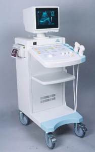 From SIUI Inc.;
'The CTS-310B is designed for the diagnosis of liver, gall, kidney, pancreas, thyroid, breast, uterus, bladder, ovary, etc. It is a versatile ultrasound scanner with both linear array scanning and convex scanning.' Features: 'Powerful image processing circuit & High quality image A wide range of probes for selection Cineloop Probe frequency conversation option Computer image communication Various measuring function Backlit keyboard'
Device Information and Specification
APPLICATIONS
See description above
CONFIGURATION
Normal system, 10' high resolution monitor, dual probe connector
Linear and convex
PROBES STANDARD
2.5MHz to 10.0MHz, linear and convex, broad band, trifrequency
IMAGING OPTIONS
Multi zoom rate and depth shift
OPTIONAL PACKAGE
POWER REQUIREMENT
AC 220V/110V, 50Hz/60Hz
POWER CONSUMPTION
0.1 KVA
• 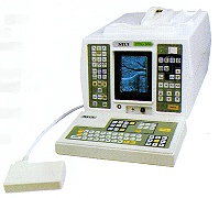 From SIUI Inc.;
'The SIUI CTS-200 is one of few 'linear only' units to be developed with a digital scan converter to process all incoming signals. With a digital processor, not only is the incoming signal processed faster but less noise is introduced into the signal path. The images are cleaner, crisper than you have experienced, even in machines at many times the price.' 'CTS-200 is suitable to a wide range of examination of liver, gallbladder, kidney, spleen, pancreas, thyroid gland, breast, uterus, urinary bladder, OB/GYN, etc. It is a portable ultrasound scanner of high performance.'
Device Information and Specification
APPLICATIONS
See description above
CONFIGURATION
Portable, gray scale(256)
Linear
PROBES STANDARD
1 * 3.5MHz Linear Probe EZU-PL21
PROBES FREQUENCY
3.5MHz, 7.5MHz
IMAGING OPTIONS
*1, *1.5, *2 as well as depth shift
OPTIONAL PACKAGE
7.5MHz Linear Probe EZU-PL23; 7.5MHz Transrectal Probe EUP-U23; 3.5MHz Biopsy Probe EUP-B11A; Trolley/Mobile Cart; ...
DATA PROCESSING
Pre-processing, correlation-processing, interpolation
POWER REQUIREMENT
AC 220V/110V, 50Hz/60Hz
POWER CONSUMPTION
0.05 KVA
•
Sound waves must have a medium to pass through. The velocity or propagation speed is the speed at which sound waves travel through a particular medium measured in meters per second (m/s) or millimeters per microsecond (mm/μs). Because the velocity of ultrasound waves is constant, the time taken for the wave to return to the probe can be used to determine the depth of the object causing the reflection. The velocity is equal to the frequency x wavelength. V = f x l The velocity of ultrasound will differ with different media. In general, the propagation speed of sound through gases is low, liquids higher and solids highest. The speed of sound depends strongly on temperature as well as the medium through which sound waves are propagating. At 0 °C (32 °F) the speed of sound in air is about 331 m/s (1,086 ft/s; 1,192 km/h; 740 mph; 643 kn), at 20 °C (68 °F) about 343 metres per second (1,125 ft/s; 1,235 km/h; 767 mph; 667 kn) Velocity (m/s)
•
air: 331;
•
fat: 1450;
•
water (50 °C): 1540;
•
human soft tissue: 1540;
•
brain: 1541;
•
liver: 1549;
•
kidney: 1561;
•
blood: 1570;
•
muscle: 1585;
•
lens of eye: 1620;
•
bone: 4080.
Doppler ultrasound visualizes blood flow-velocity information. The peak systolic velocity and the end diastolic velocity are major Doppler parameters, which are determined from the spectrum obtained at the point of maximal vessel narrowing. Peak systolic velocity ratios are calculated by dividing the peak-systolic velocity measured at the site of flow disturbance by that measured proximal of the narrowing (stenosis, graft, etc.). See Acceleration Index, Acceleration Time, Modal Velocity, Run-time Artifact and Maximum Velocity. Further Reading: Basics:
News & More:
•
Vascular ultrasound contrast agents are gas microbubbles with a diameter less than 10 μm (2 to 5 μm on average for most of the newer agents) to pass through the lung capillaries and enter into the systemic circulation. Air bubbles in that size persist in solution for only a short time; too short for systemic vascular use in medical ultrasound imaging. So the gas bubbles have to be stabilized to persist long enough and survive pressure changes in the heart. Most vascular contrast media are stabilized against dissolution and coalescence by the presence of additional materials at the gas-liquid interface. In some cases, this material is an elastic solid shell that enhances stability by supporting a strain to counter the effect of surface tension. In other cases, the material is a surfactant, or a combination of two or more surfactants. Typically the effective duration of vascular enhancement is a few minutes, after which the microbubbles dissipate. This rather short duration of vascular enhancement makes it easy to perform repeated dynamic studies. Intravenous vascular contrast agents will be used in imaging malignant tumors in the liver, kidney, ovary, pancreas, prostate, and breast. Tumor neovascularization can be a marker for angiogenesis, and Doppler signals from small tumor vessels may be detectable after contrast injection. Contrast agents are useful for evaluating vessels in a variety of organs, including those involved in renal, hepatic, and pancreatic transplants. If an area of ischemia or a stenosis is detected after contrast administration, the use of other more expensive imaging modalities, including CT and MRI, can often be avoided. See also Acoustically Active Lipospheres. Result Pages : | Share This Page Look Ups |
Medical-Ultrasound-Imaging.com
former US-TIP.com
Member of SoftWays' Medical Imaging Group - MR-TIP • Radiology TIP • Medical-Ultrasound-Imaging
Copyright © 2008 - 2024 SoftWays. All rights reserved.
Terms of Use | Privacy Policy | Advertise With Us
former US-TIP.com
Member of SoftWays' Medical Imaging Group - MR-TIP • Radiology TIP • Medical-Ultrasound-Imaging
Copyright © 2008 - 2024 SoftWays. All rights reserved.
Terms of Use | Privacy Policy | Advertise With Us
[last update: 2023-11-06 01:42:00]