Medical Ultrasound Imaging
Thursday, 9 May 2024
Info Sheets Out- side     | 'Out of Phase' p4 Searchterm 'Out of Phase' found in 19 articles 1 definition [ • ] - 18 booleans [• ]Result Pages : • 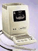 From Philips Medical Systems;
'The perfect practice system... The SD 240E is a compact and ergonomically designed system, standard equipped with two probe connectors. The user has a choice out of more than 12 linear, curved and annular phased array probes which can be used with this perfect office unit. Total Image Focus and cine memory technology are just the icing on the cake.' This product is not available in the US, Australia, or New Zealand from Philips Medical Systems.' Specifications for this system will be available soon. • 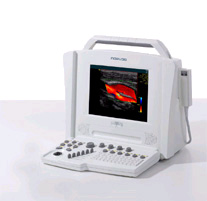 From Siemens Medical Systems;
We see a way to review 100% of your cardiovascular ultrasound exams and reports anywhere, anytime. The Cypress™ system is a highly miniaturized, all digital, phased array echocardiography system that provides complete studies and outstanding images - even on the most technically difficult patients. Specifications for this system will be available soon. •
Standard scanners allow visualizing microbubbles on conventional gray scale imaging in large vascular spaces. In the periphery, more sensitive techniques such as Doppler or non-linear gray scale modes must be used because of the dilution of the microbubbles in the blood pool. Harmonic power Doppler (HPD) is one of the most sensitive techniques for detecting ultrasound contrast agents. Commonly microbubbles are encapsulated or otherwise stabilized to prolong their lifetime after injection. These bubbles can be altered by exposure to ultrasound pulses. Depending on the contrast agent and the insonating pulse, the changes include deformation or breakage of the encapsulating or stabilizing material, generation of free gas bubbles, reshaping or resizing of gas volumes. High acoustic pressure amplitudes and long pulses increase the changes. However, safety considerations limit the pressure amplitude and long pulses decrease spatial resolution. In addition, lowering the pulse frequency increases destruction of contrast bubbles. However, at low insonation power levels, contrast agent particles resist insonation without detectable changes. Newer agents are more reflective and will usually allow gray scale imaging to be used with the advantages of better spatial resolution, fewer artifacts and faster frame rates. Feasible imaging methods with advantages in specific acoustic microbubble properties: Resonating microbubbles emit harmonic signals at double their resonance frequency. If a scanner is modified to select only these harmonic signals, this non-linear mode produces a clear image or trace. The effect depends on the fact that it is easier to expand a bubble than to compress it so that it responds asymmetrically to a symmetrical ultrasound wave. A special array design allows to perform third or fourth harmonic imaging. This probe type is called a dual frequency phased array transducer. See also Bubble Specific Imaging. •
Ultrasound machines, widely used in medical imaging, are essential tools in the field of diagnostic ultrasound. These devices utilize high-frequency sound waves to create real-time images of internal body structures. Ultrasound machines consist of several key components that work together to generate diagnostic images.
These include:
•
The transducer is a handheld device that emits and receives sound waves. It converts electrical energy into sound waves and captures the returning echoes to create images.
•
The control panel houses the interface where the sonographer adjusts imaging parameters such as depth, frequency, and gain. It allows for customization of imaging settings based on the clinical requirements. The transducer pulse controls change the amplitude, frequency and duration of the pulses emitted from the transducer probe.
•
The central processing unit (CPU) serves as the brain of the ultrasound machine, processing the acquired data and transforming it into images. It handles complex calculations, image optimization, data storage and contains the electrical power supplies for itself and the transducer probe.
•
The display monitor (oscilloscope, tablet, computer monitor, etc.) showcases the real-time ultrasound images produced by the machine. It provides visual feedback to the sonographer, aiding in the interpretation and analysis of anatomical structures. Handheld ultrasound devices and mobile ultrasound probes can be connected wirelessly to a smartphone or tablet via Bluetooth or WiFi. These end device serves then as the ultrasound monitor.
•
Data input and measurements are done with the keyboard cursor (trackball). Ultrasound devices used for handheld point of care ultrasound (HPOCUS) are operated via the touch screen of the control panel.
•
Images are captured, reviewed, stored and transmitted digitally, using a standard format for digital imaging and communications in medicine (DICOM). Disk storage devices (FDD, HDD, CD, DVD) are outdated, but may be used in older machines to store the acquired images if no picture archiving and communication system (PACS) connection is possible.
•
The displayed ultrasound pictures are usually digitally stored in a PACS. The images from portable ultrasound machines can be stored and conveniently managed on the end device itself, the inserted memory card or in the cloud. With a QR scanner, the images can be accessed via the Internet in the cloud. Often there is also the possibility to get a picture of a baby sonography as a printout.
B-mode machines represent the vast majority of machines used in echocardiology, obstetrical scans, abdominal scans, gynecological scans, etc. B-mode ultrasound machines usually produce the sector (or pie segment-shaped) scans. These ultrasound scans require either a mechanical scanner transducer (the transducer moves to produce the sector scan), or a linear array transducer operated as a phased array. Ultrasound machines come in different types, each catering to specific clinical needs. The two primary types are stationary and portable ultrasound machines: •
Stationary units are typically larger in size and are installed in dedicated imaging rooms. These machines offer advanced imaging capabilities and a wide range of specialized features. They are commonly found in hospitals, clinics, and university medical centers where comprehensive imaging services are provided.
•
Portable units (see Portable Ultrasound Machine), as the name suggests, are compact and lightweight, designed for on-the-go imaging. These machines are highly versatile and offer excellent mobility, allowing healthcare professionals to bring the ultrasound system directly to the patient's bedside. Portable ultrasound machines are particularly useful in emergency settings, rural healthcare facilities, and point-of-care applications.
See also Handheld Ultrasound, Ultrasound System Performance, Equipment Preparation, Coaxial Cable, and Microbubble Scanner Modification, Environmental Protection and Ultrasound Accessories and Supplies. Further Reading: Basics: News & More:
Result Pages : | Share This Page Look Ups |
Medical-Ultrasound-Imaging.com
former US-TIP.com
Member of SoftWays' Medical Imaging Group - MR-TIP • Radiology TIP • Medical-Ultrasound-Imaging
Copyright © 2008 - 2024 SoftWays. All rights reserved.
Terms of Use | Privacy Policy | Advertise With Us
former US-TIP.com
Member of SoftWays' Medical Imaging Group - MR-TIP • Radiology TIP • Medical-Ultrasound-Imaging
Copyright © 2008 - 2024 SoftWays. All rights reserved.
Terms of Use | Privacy Policy | Advertise With Us
[last update: 2023-11-06 01:42:00]