Medical Ultrasound Imaging
Saturday, 18 May 2024
Info Sheets Out- side     | 'Phase' p3 Searchterm 'Phase' found in 77 articles 4 terms [ • ] - 73 definitions [• ] Result Pages : •
[This entry is marked for removal.] From POINT Biomedical Corp CARDIOsphere® is an ultrasound contrast agent for assessment of myocardial perfusion in patients with coronary artery disease composed of highly echogenic bispheres. PB127 is a new developed microbubble with a bilayer polymer/albumin shell filled with nitrogen gas that has ideal characteristics for power harmonic Doppler. They can be destroyed by high power ultrasound, and spectral decorrelation between ultrasound pulses is maximized by rapid dissolution of the released nitrogen gas. POINT Biomedical Corp. announced (March 01, 2004) that it has completed two Phase 3 trials of CARDIOsphere®. The Phase 3 trials were designed to evaluate the performance of CARDIOsphere® imaging relative to radionuclide imaging for detecting obstructive coronary artery disease and identifying the anatomic location of perfusion defects.
Drug Information and Specification
RESEARCH NAME
PB 127
DEVELOPER
INDICATION -
DEVELOPMENT STAGE APPLICATION
Infusion
TYPE
Microbubble
Polylactide/Albumin
CHARGE
Slight Negative
Nitrogen
PREPARATION
Reconstitute with 2ml H2O per vial and dilute with 150 ml DSW
DO NOT RELY ON THE INFORMATION PROVIDED HERE, THEY ARE
NOT A SUBSTITUTE FOR THE ACCOMPANYING PACKAGE INSERT! •
Quadrature detection is used in Doppler ultrasound as well as in magnetic resonance imaging and is also called quadrature demodulation or phase quadrature technique. Quadrature detection is the acquisition of Mx and My simultaneously as a function of time by using two separate detector channels. This signal processing method is used for directional Doppler in which the signal reference frequency for the two channels has a phase shift of 1/4 period. The output Doppler signal phase for both channels also depends on the Doppler shift whether positive or negative. The fast Fourier transform analyzer performs spectral Doppler analysis in ultrasound machines and displays different quadrature Doppler frequencies, when a sample volume cursor is used along time. • 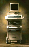 From SIUI Inc.;
'The Apogee 800Plus ultrasound system offers a new level of high quality in image. With broad clinical application, the system delivers ease of use for cardiac and vascular exams.' 'Due to the exceptional image quality, the sensitivity of color Doppler and many other advanced features, the Apogee 800Plus offers complete diagnosis capability for your entire patient population, including abdominal, obstetrics, gynecology, adult cardiology, pediatric cardiology, peripheral vascular, small parts, breast, prostate, transcranial, etc.'
Device Information and Specification
APPLICATIONS
Linear, curved, phased and symmetric phased array
2D, M-mode, Pulsed wave Doppler, Color Power Angio Imaging, Color 2D, Color M-mode, High PRF Doppler
H*W*D m
1.46 * 0.69 * 0.87
WEIGHT
140 kg (without peripherals)
POWER REQUIREMENT
115Vac, (V~), @ 50 to 60 Hz, 1.2kVA
230Vac, (V~), @ 50 to 60 Hz, 1.2kVA POWER CONSUMPTION
800 Watts (with optional OEM'S: 1200 Watts max)
•
Bubble specific imaging methods rely usually on non-linear imaging modes. These contrast imaging techniques are designed to suppress the echo from tissue in relation to that from a microbubble contrast agent. Stimulated acoustic emission (SAE) and phase / pulse inversion imaging mode (PIM) are bubble specific modes, which can image the tissue specific phase. In SAE mode bubble rupture is seen as a transient bright signal in B-mode and as a characteristic mosaic-like effect in velocity 2D color Doppler. PIM are Doppler modes and detect non-linear echoes from microbubbles. In pulse inversion imaging modes the transducer bandwidth extends, resulting in improved spatial resolution and more contrast. See also Contrast Pulse Sequencing, Microbubble Scanner Modification, Narrow Bandwidth, Contrast Medium, Dead Zone. Further Reading: Basics:
Result Pages : | Share This Page Look Ups |
Medical-Ultrasound-Imaging.com
former US-TIP.com
Member of SoftWays' Medical Imaging Group - MR-TIP • Radiology TIP • Medical-Ultrasound-Imaging
Copyright © 2008 - 2024 SoftWays. All rights reserved.
Terms of Use | Privacy Policy | Advertise With Us
former US-TIP.com
Member of SoftWays' Medical Imaging Group - MR-TIP • Radiology TIP • Medical-Ultrasound-Imaging
Copyright © 2008 - 2024 SoftWays. All rights reserved.
Terms of Use | Privacy Policy | Advertise With Us
[last update: 2023-11-06 01:42:00]