Medical Ultrasound Imaging
Sunday, 19 May 2024
Info Sheets Out- side     | 'Radio Frequency' Searchterm 'Radio Frequency' found in 7 articles 2 definitions [ • ] - 5 booleans [• ]Result Pages : •
(IVUS) For intravascular ultrasound a small IVUS catheter with a probe is introduced into the artery. The transducer transmits and receives acoustic energy through this catheter. The reflected acoustic energy is used to build a picture of the inside of the vessel. A IVUS image consists of three layers around the lumen, the intima, media and adventitia. In addition, elastography or palpography could be used to evaluate the local mechanical properties of tissues (e.g. lipid pools in high-risk vulnerable atherosclerotic plaques). These techniques use the deformation caused by the intraluminal pressure generated by the probe. A high strain region at the lumen vessel wall boundary has 88% sensitivity and 89% specificity for identifying vulnerable plaques. There are high strain values of 1% in soft plaques with increased strain up to 2% at the shoulders of the plaque, while calcified material shows low strain values (0-0.2%). The radial strain in the tissue is obtained by cross-correlation techniques on the radio frequency signal. The strain is color-coded and plotted as a complimentary image to the intravascular ultrasound echogram. See also Interventional Ultrasound, Vascular Ultrasound. • View NEWS results for 'Intravascular Ultrasound' (2). •
Radio frequency (RF) thermal ablation is a technique that uses the thermal effect created by radio frequencies to destroy tumors or metastases in the liver. This treatment for liver cancer can accurately be evaluated by contrast enhanced ultrasound. RF thermal ablation monitored by sonography can lead to immediate re-treatment, preventing a second anesthesia and shortening the hospitalization time. Further Reading: Basics: • 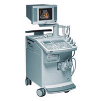 From Siemens Medical Systems;
'This extremely flexible system supports targeted applications for OB/GYN, Radiology, Internal Medicine, Urology, and Prostate Brachytherapy. The large transducer selection, most with high-frequency capabilities, provides the right tool for general imaging, radiology, and internal medicine applications.'
Device Information and Specification
CLINICAL APPLICATION
OB/GYN, cerebrovascular, orthopedics, urology, peripheral vascular, pediatrics, small parts, surgery, abdominal, musculoskeletal
CONFIGURATION
Mobile, compact
DISPLAY MODES
Broadband, high-fidelity, multi-frequency, linear and curved
PROBE PORTS
Two - four
256
MEASUREMENT/CALCULATION FUNCTIONS
OB/GYN measurements and report package, off-line analysis
OPTIONAL PACKAGE
IMAGE PROCESSING
•
(LUS) Diagnostic laparoscopy combined with laparoscopic ultrasound is used for staging tumors and to monitor surgical interventions like for example radiofrequency ablation or cryotherapy. Laparoscopic ultrasound provides direct contact imaging of organs with high frequency ultrasound. Laparoscopic ultrasound identifies and characterizes the tumor, guides the probe, and monitors the progression of the freezing or the thermal destruction. This procedure avoid unnecessary open surgery and improves selection of patients for tumor resection e.g., in liver and pancreas. Challenges of LUS are limitations of the intraoperative acoustic windows and the possible movement of the probe and that standard orientation techniques are difficult to apply with laparoscopic instruments, resulting in images from oblique planes. 3D ultrasound or special navigation systems may be helpful. See also Ultrasound Therapy. •
The wavelength is a unit of relative distance equal to the length of a wave. This could be a light wave, a radio wave, or even a sound wave.
For sound waves the formula is: l=c/f (wavelength = propagation speed/frequency) In ultrasound imaging is the wavelength the distance between the onset of peak compression or cycle to the next. The wave propagates as bands of compression and rarefaction. One wavelength is the distance between two bands of compression, or rarefaction. Maximum compression corresponds to maximum pressure. The wavelength (see also Angstrom) is important in image resolution. See also Spectral Reflector. Result Pages : | Share This Page Look Ups |
Medical-Ultrasound-Imaging.com
former US-TIP.com
Member of SoftWays' Medical Imaging Group - MR-TIP • Radiology TIP • Medical-Ultrasound-Imaging
Copyright © 2008 - 2024 SoftWays. All rights reserved.
Terms of Use | Privacy Policy | Advertise With Us
former US-TIP.com
Member of SoftWays' Medical Imaging Group - MR-TIP • Radiology TIP • Medical-Ultrasound-Imaging
Copyright © 2008 - 2024 SoftWays. All rights reserved.
Terms of Use | Privacy Policy | Advertise With Us
[last update: 2023-11-06 01:42:00]