Medical Ultrasound Imaging
Sunday, 19 May 2024
Info Sheets Out- side     | 'Rectal Probe' p3 Searchterm 'Rectal Probe' found in 23 articles 1 term [ • ] - 16 definitions [• ] - 6 booleans [• ]Result Pages : • 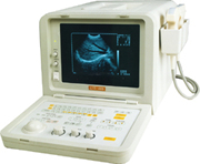 From SIUI Inc.;
'SIUI's newly developed CTS-485 is a portable ultrasound system with advanced technology. With its powerful features, such as 256 grayscale, cineloop, RS232C interface and broadband high-density, multi-frequency probes, the system is designed for professional applications in cardiology, abdominal, small parts, liver, gallbladder, kidney, obstetrics, gynecology, peripheral vascular, etc. The CTS-485 has passed FDA clearance and CE Marking.'
Device Information and Specification
APPLICATIONS
CONFIGURATION
Portable, gray scale(256)
Linear and convex
PROBES STANDARD
1 * 2.5MHz ~ 5.0MHz multifrequency convex probe
2.5MHz to 10.0MHz broad band, trifrequency
IMAGING OPTIONS
Multi zoom rate and depth shift
OPTIONAL PACKAGE
H*W*D m
0.26 * 0.3 * 0.41
WEIGHT
11 kg (main unit)
POWER REQUIREMENT
AC 220V/110V, 50Hz/60Hz
POWER CONSUMPTION
0.1 KVA
•  From SIUI Inc.;
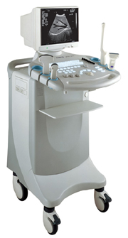
'Incorporating the latest advances in technology, the CTS-6000 digital B/W ultrasound system provides exceptional imaging quality and complete diagnostic capabilities. It is equipped with extensive analysis package, super broadband multifrequency probes and disk storage capacity, a complete system that fully meets different clinical needs. CTS-6000 sets a new standard for digital ultrasound imaging.'
Device Information and Specification
CONFIGURATION
Normal system, 12-inch high-resolution non-interlace monitor , Tri-probe connector
Linear and convex
PROBES STANDARD
1 * Super broadband linear probe L7S34, 1 * super broadband micro-convex probe C3L60, 1 * super broadband micro-convex probe C3120;
IMAGING OPTIONS
Real ZOOM, max. zoomx4.0, position selectable
H*W*D m
1.28 * 0.48 * 0.72
WEIGHT
98kg (main unit)
POWER REQUIREMENT
AC 220V/110V, 50Hz/60Hz
POWER CONSUMPTION
0.43 KVA
•
The usual applications of endocavitary echography (also called internal echography / endoscopic ultrasound (EUS)) are examinations of the pelvic organs through internally introduced probes, which give a more precise and correct image. Transrectal ultrasound is a well established method for rectal or prostate carcinoma assessment. A transvaginal echography uses a small transducer that is inserted directly into the vagina. Used are high-frequency (10-12 MHz) for superficial organs, endocavitary echography, and intraoperative laparoscopic ultrasound. A sterile cover is slipped over the probe, which is then covered with lubricating ultrasound gel and placed in the cavitary (see Equipment Preparation). See also Endoscopic Ultrasound, Prostate Ultrasound, Interventional Ultrasound, Transurethral Sonography, Vaginal Probe, Rectal Probe. •
The prostate is a walnut-shaped gland surrounding the beginning of the urethra in front of the rectum and below the bladder. The prostate can become enlarged (particularly in men over age 50) and develop diseases like prostate cancer or inflammation (prostatitis). A large tumor can be felt by a rectal examination. The most effective way of detecting the early signs of prostate cancer is a combination of a prostate-specific antigen (PSA) blood test and a prostate ultrasound examination. An abnormally high level of PSA can indicate prostate cancer or other prostate diseases such as benign prostatic hypertrophy or prostatitis. The transrectal sonography is an important diagnostic ultrasound procedure in determining whether there is any benign enlargement of the prostate or any abnormal nodules. The imaging is performed with a rectal probe, yielding high resolution. High resolution 3D ultrasound provides reliable and accurate determination of the size and the location of cancer. Additionally, ultrasound elastography is a technique in development to improve the specificity and sensitivity of cancer detection. Ultrasound is also used to detect whether cancerous tissue is still only within the prostate or whether it has begun to spread out and to guide a diagnostic biopsy or ultrasound therapy. See also Brachytherapy, and High Intensity Focused Ultrasound. Further Reading: News & More:
•
A transducer assembly is the configuration of an ultrasound transducer. Electronic array transducers are composed of multiple crystal elements e.g., linear array and phased array transducer assemblies. A handheld mechanical probe may contain a mechanically driven single element transducer, or a rotating wheel transducer. Static B-scanner (obsolete) had a cylindrical probe with a single disk-shaped piezoelectric crystal. Some probes are designed to be inserted in body cavities (vagina, rectum, esophagus) so that they can get closer to the organ being examined (uterus, prostate gland, heart); getting closer to the region of interest allow for a more detailed view. See also Endocavitary Echography, Endoscopic Ultrasound, Vaginal Transducer and Rectal Probe. Result Pages : | Share This Page Look Ups |
Medical-Ultrasound-Imaging.com
former US-TIP.com
Member of SoftWays' Medical Imaging Group - MR-TIP • Radiology TIP • Medical-Ultrasound-Imaging
Copyright © 2008 - 2024 SoftWays. All rights reserved.
Terms of Use | Privacy Policy | Advertise With Us
former US-TIP.com
Member of SoftWays' Medical Imaging Group - MR-TIP • Radiology TIP • Medical-Ultrasound-Imaging
Copyright © 2008 - 2024 SoftWays. All rights reserved.
Terms of Use | Privacy Policy | Advertise With Us
[last update: 2023-11-06 01:42:00]