Medical Ultrasound Imaging
Friday, 10 May 2024
Info Sheets Out- side     | 'Resolution' p2 Searchterm 'Resolution' found in 71 articles 5 terms [ • ] - 66 definitions [• ] Result Pages : •
Ultrasound machines, with their various components and types, have revolutionized the field of medical imaging. These devices enable healthcare professionals to visualize internal structures, assess conditions, and guide interventions with real-time imaging capabilities.
Today, medical ultrasound systems are complex signal processing machines. Assessing the performance of an ultrasound system requires understanding the relationships between the characteristics of the system, such as the point spread function, temporal resolution, and the quality of images. Image quality aspects include the detail resolution, contrast resolution and penetration. Systems with microbubble scanner modification are particularly suitable for contrast enhanced ultrasound.
•
Low-performance systems constitute approximately 20% of the world ultrasound market. These ultrasound machines are characterized by basic black and white imaging and are primarily used for basic OB/GYN applications and fetal development monitoring. They are often purchased by private office practitioners and small hospitals, with a unit cost below $50,000. These scanners commonly come equipped with a transvaginal probe.
•
Mid-performance sonography systems also hold around 20% market share. These machines are basic gray scale imaging, color and spectral Doppler and are used for routine examinations and reporting. They typically utilize a minimum number of scanheads and find applications in radiology, cardiology, and OB/GYN. The cost of these systems ranges between $50,000 and $100,000. Refurbished advanced and high-performance ultrasound machines with fewer optional features can also be found in this price range.
•
High-performance ultrasound systems generally provide high-resolution gray scale imaging, advanced color power and spectral Doppler capabilities. They usually include advanced measurement and analysis software, image review capabilities, and a variety of probes. These high-performance sonography devices have a market share of approximately 40% and cost between $100,000 and $150,000.
•
The remaining 20% of the market consists of premium or advanced performance ultrasound systems, typically sold for over $150,000. Premium performance systems offer high-resolution gray scale imaging, advanced color flow, power Doppler, and spectral Doppler, as well as features like tissue harmonic imaging, image acquisition storage, display and review capabilities, advanced automation, and more. Premium systems are equipped with a wide assortment of transducer scanheads.
In summary, ultrasound machines have diverse performance levels and corresponding price ranges, catering to various medical imaging needs. From low-performance systems with basic imaging capabilities to high-performance and premium systems with advanced features, ultrasound technology continues to advance healthcare imaging capabilities. See also Ultrasound Physics, Handheld Ultrasound, Environmental Protection, Equipment Preparation. Further Reading: Basics:
•
(THI) Tissue harmonic imaging (also called native harmonic imaging) is a signal processing technique which addresses ultrasound limitations like penetration and resolution. Tissue harmonic imaging reduces noise and clutter by improving signal to noise ratio and resolution. The signal penetration in soft tissue increases as the transmit frequency is decreased, by simultaneous decreased image resolution. As an ultrasound wave propagates through the target media a change occurs in the shape and frequency of the transmitted signal. The change is due to the normal resistance of tissue to propagate sound energy. This resistance and the resulting signal change is called a harmonic oscillation. For harmonic imaging the input frequency doubles the output frequency, for example a transmit frequency of 3.0 MHz. which would provide maximum penetration will return a harmonic frequency of 6.0 MHz. The returning higher frequency signal has to only travel one direction to the probe. The advantages of high frequency imaging and the one-way travel effect are decreased reverberation, beam aberration, and side lobes, as well as increased resolution and cystic clearing. •
Ultrasound biomicroscopy utilizes high frequency (10 - 50 MHz) diagnostic ultrasound to examine living tissue at a microscopic level and allows to image the skin with extremely high resolution to a depth of 2-3 centimeters. Ultrasound biomicroscopy images provide detailed anatomical information that can lead to better and more accurate treatments and avoid a biopsy. Ultrasound biomicroscopy improves also the spatial resolution of US images of the anterior segment of the eye. US biomicroscopy of the eye operates in the 50 MHz range with a possible axial resolution on the order of 30 μm. In this frequency range, tissue penetration of only approximately 5 mm is attainable. Both continuous wave Doppler and high-frequency pulsed Doppler can be used. See also Ultrasound Imaging Procedures, A-Scan, B-Scan and C-Scan. Further Reading: News & More:
•
A phantom is used to control the imaging performance of ultrasound transducers. The spatial resolution, dead zone, linear fidelity, depth of penetration and image uniformity is important for the image quality. For the axial and lateral resolution, the standard definition is the resolution of objects parallel and perpendicular to the path of the sound beam. Ultrasound pictures created by scans of specially designed ultrasound phantoms can quantify the imaging quality and transducer performance. Phantoms contain one or more materials that simulate a tissue in its interaction with ultrasound. Several phantoms are available specifically for quality control. Transducer characterization consists of a standard pulse echo analysis and insertion loss measurement for each probe. The quality variation from the baseline should be tracked over a period. Further Reading: Basics: • 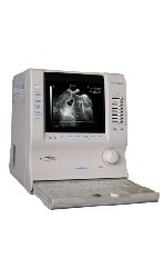 From ALOKA Co., Ltd.;
'Innovative Image Quality in a Compact Size. Featuring design concepts inherited from our higher end systems along with Aloka's latest proprietary electronic technologies, the compact SSD-900 delivers superb images. Traditionally, portable units suffer from hardware and software limitations that affect image quality. Aloka's SSD-900 sets new standards for what's possible in a portable unit. Incorporating over 50 years of experience and unique micro-chip technologies, the SSD-900 generates incredibly high-resolution images. The SSD-900 offers a full range of measurement functions for professional ultrasonic examination. And unlike conventional portable systems, the SSD-900 includes annotation labeling and 15-channel preset functions as standard features. The system also use Super High Density transducers (found our high-end systems) to enhance imaging resolution. These multi-frequency transducers provide a broad range of imaging frequencies to optimize scan frequency for depth of view.' Result Pages : | Share This Page Look Ups |
Medical-Ultrasound-Imaging.com
former US-TIP.com
Member of SoftWays' Medical Imaging Group - MR-TIP • Radiology TIP • Medical-Ultrasound-Imaging
Copyright © 2008 - 2024 SoftWays. All rights reserved.
Terms of Use | Privacy Policy | Advertise With Us
former US-TIP.com
Member of SoftWays' Medical Imaging Group - MR-TIP • Radiology TIP • Medical-Ultrasound-Imaging
Copyright © 2008 - 2024 SoftWays. All rights reserved.
Terms of Use | Privacy Policy | Advertise With Us
[last update: 2023-11-06 01:42:00]