Medical Ultrasound Imaging
Friday, 10 May 2024
Info Sheets Out- side     | 'Resolution' p3 Searchterm 'Resolution' found in 71 articles 5 terms [ • ] - 66 definitions [• ] Result Pages : • 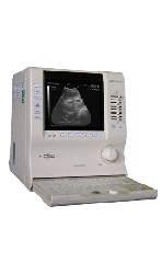 From ALOKA Co., Ltd.;
'Innovative Image Quality in a Compact Size Featuring design concepts inherited from our higher end systems along with Aloka's latest proprietary electronic technologies, the compact SSD-900V delivers superb images. Traditionally, portable units suffer from hardware and software limitations that affect image quality. Aloka's SSD-900V sets new standards for what's possible in a portable unit. Incorporating over 50 years of experience and unique micro-chip technologies, the SSD-900V generates incredibly high-resolution images. The SSD-900V offers a full range of measurement functions for professional ultrasonic examination. And unlike conventional portable systems, the SSD-900V includes annotation labeling and 15-channel preset functions as standard features. The system also use Super High Density transducers (found our high-end systems) to enhance imaging resolution. These multi-frequency transducers provide a broad range of imaging frequencies to optimize scan frequency for depth of view.' •
The wider the ultrasound beam, the more severe the problem with volume averaging and the beam-width artifact, to avoid this, the ultrasound beam can be shaped with lenses.
Different possibilities to focus the beam:
•
Mechanical focusing is performed by placing an acoustic lens on the surface of the transducer or using a transducer with a concave face.
•
Electronic focusing uses multiple phased array (annular or linear) elements, sequentially fired to focus the beam.
Conventional multi-element transducers are electronically focused in order to minimize beam width. This transducer type can be focused electronically only along the long axis of the probe where there are multiple elements, along the short axis (elevation axis) are conventional transducers only one element wide. Electronic focusing in any axis requires multiple transducer elements arrayed along that axis. Short axis focusing of conventional multi-element transducers requires an acoustic lens which has a fixed focal length. For operation at frequencies at or even above 10 MHz, quantization noise reduces contrast resolution. Digital beamforming gives better control over time delay quantization errors. In digital beamformers the delay accuracy is improved, thus allowing higher frequency operation. In analog beamformers, delay accuracy is in the order of 20 ns. Phased beamformers are suitable to handle linear phased arrays and are used for sector formats such as required in cardiography to improve image quality. Beamforming in ultrasound instruments for medical imaging uses analog delay lines. The signal from each individual element is delayed in order to steer the beam in the desired direction and focuses the beam. The receive beamformer tracks the depth and focuses the receive beam as the depth increases for each transmitted pulse. The receive aperture increase with depth. The lateral resolution is constant with depth, and decreases the sensitivity to aberrations in the imaged tissue. A requirement for dynamic control of the used elements is given. Since often a weighting function (apodization) is used for side lobe reduction, the element weights also have to be dynamically updated with depth. See also Huygens Principle. •
Breast ultrasound (sonography or ultrasonography) it is an important tool in the characterization of breast lesions, detected with mammography or clinical breast examination. However, a breast sonogram is not approved by the U.S. Food and Drug Administration (FDA) as a screening tool for breast cancer and is used additional to a mammogram. Ultrasound is useful in guiding needles for fine needle aspiration and core biopsies. Breast ultrasound has optimal contrast resolution, but it lacks the spatial resolution of conventional mammography and cannot provide as much detail as a mammogram image. In addition, ultrasound is unable to show tiny calcium deposits (microcalcifications) that are often early indications of breast cancer. See also Biopsy, Interventional Ultrasound, Ultrasound Safety, Side Effect and Ultrasound Regulations. •
Increasing the frequency of the transmitted power improves the image quality of ultrasound, but the improvement in resolution results in a decreased signal to noise ratio (SNR). Higher acoustic power levels can prevent the loss in SNR, but among other reasons, ultrasound regulations limit this to avoid heating or cavitation. Coded excitation increase the signal to noise ratio without the loss of resolution by using coded waveforms. Coded excitation allows transmitting a long wide-band pulse with more acoustic power and high penetration of the sound beam. •
Damping is a process, material, design, and mounting technique used to reduce the pulse duration or ringing of the transducer. Special material is applied to the back of the transducer in order to reduce the amplitude and pulse length of the sound wave. Damping improves axial resolution by reducing pulse length. Thereby the lateral resolution increases. Result Pages : | Share This Page Look Ups |
Medical-Ultrasound-Imaging.com
former US-TIP.com
Member of SoftWays' Medical Imaging Group - MR-TIP • Radiology TIP • Medical-Ultrasound-Imaging
Copyright © 2008 - 2024 SoftWays. All rights reserved.
Terms of Use | Privacy Policy | Advertise With Us
former US-TIP.com
Member of SoftWays' Medical Imaging Group - MR-TIP • Radiology TIP • Medical-Ultrasound-Imaging
Copyright © 2008 - 2024 SoftWays. All rights reserved.
Terms of Use | Privacy Policy | Advertise With Us
[last update: 2023-11-06 01:42:00]