Medical Ultrasound Imaging
Sunday, 19 May 2024
Info Sheets Out- side     | 'Ultrasound Technology' p3 Searchterm 'Ultrasound Technology' found in 36 articles 1 term [ • ] - 18 definitions [• ] - 17 booleans [• ]Result Pages : • 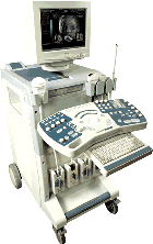 From Medison Co.,Ltd.;
'The SONOACE 9900 offers exceptional B/W image clarity by newly developed FINE™ Filter technology, which contributes to edge enhancement and average filtering in real-time. OSIO™(Organ Specified Image Optimization)sets the best diagnosis environment with minimum manual operation and increases patient throughput. Also, color images are improved with better color Doppler performance, high color sensitivity, fine pixel image, and reduced flash artifact across all probes.' 'The SONOACE 9900 PRIME from Medison offers full range of applications specially designed for your practice. With the addition of the new C-Square Processing and PSAD Beamformer, SONOACE 9900 PRIME provides higher contrast resolution and clearer image quality in all application ranges. Based on super clear image resolution and versatile 3D functions, SONOACE 9900 PRIME offers you the true value of future ultrasound technology.' •
The term 'sonogram' is often used interchangeably with 'ultrasound,' but it specifically refers to the resulting image or picture produced during a diagnostic ultrasound examination, also known as ultrasonography or sonography. It serves as a visual representation of the echoes detected by the transducer and provides detailed anatomical information about the area being examined. Sonograms are typically displayed on a monitor, printed on film, or stored digitally for further analysis and documentation by medical professionals such as sonographers and radiologists. They serve as invaluable diagnostic tools, aiding in the detection and evaluation of various medical conditions, as well as guiding interventions, ultrasound therapy, and treatment planning. The term 'ultrasound' itself refers to the technology used during a sonogram, but it also finds several other applications beyond medical imaging. These include echolocation, crack detection, and cleaning, among others. See also Ultrasound Imaging, Ultrasound Technology, Handheld Ultrasound, Ultrasound Accessories and Supplies, Environmental Protection and Ultrasound Elastography. •
Sonography [aka: ultrasonography] is a term that encompasses the entire process of performing ultrasound examinations and interpreting the obtained images. Sonography involves the skilled application of ultrasound technology by trained professionals known as sonographers or ultrasound technologists. These specialists operate the ultrasound equipment, manipulate the transducer, and acquire the necessary pictures for diagnostic imaging purposes. Sonography requires in-depth knowledge of anatomy, physiology, and pathology to accurately interpret the ultrasound images and provide valuable information to the treating physician. Sonography uses equipment that generates high frequency sound waves to produce images from muscles, soft tissues, fluid collections, and vascular structures of the human body. Obstetric sonography is commonly used during pregnancy. Sonography visualizes anatomy, function, and pathology of for example gallbladder, kidneys, pancreas, spleen, liver, uterus, ovaries, urinary bladder, eye, thyroid, breast, aorta, veins and arteries in the extremities, carotid arteries in the neck, as well as the heart. A typical medical ultrasound machine, usually a real-time scanner, operates in the frequency range of 2 to 13 megahertz. See also Musculoskeletal and Joint Ultrasound, Pediatric Ultrasound, Cerebrovascular Ultrasonography and Contrast Enhanced Ultrasound. Further Reading: Basics:
News & More:
•
Ultrasonography is another term[aka: sonography] used to describe the practice of using ultrasound technology for diagnostic imaging. It is synonymous with sonography and signifies the process of capturing ultrasound images, regardless of the body part or condition being examined. Ultrasonography is widely utilized in various medical imaging specialties, including obstetrics and gynecology, cardiology, radiology, urology, and many others. It has proven to be particularly valuable in obstetric imaging, allowing healthcare providers to monitor the growth and development of a fetus during pregnancy.
Ultrasonography uses the reflections of high-frequency sound waves to construct an image of a body organ. These ultrasonic waves are generated by a quartz crystal and are reflected at the interface between different tissues. The transmission and reflection of these high-frequency waves are displayed with different types of ultrasound modes. See also sonogram, sonography, ultrasound imaging. Further Reading: News & More:
•
Common ultrasound supplies that are often used in conjunction with ultrasound imaging:
•
Ultrasound Gel: A water-based gel used as a coupling agent between the transducer and the patient's skin. It helps eliminate air pockets and ensures good sound wave transmission. •
Probe Covers: Disposable covers designed to maintain hygiene and prevent cross-contamination. These covers are placed over the transducer before each examination. •
Cleaning Wipes: Alcohol-based or disinfectant wipes used for cleaning and disinfecting the transducer and other equipment surfaces. Specific cleaning solutions are recommended by the ultrasound equipment manufacturer for thorough cleaning of transducers. •
Gel Warmers: Devices used to warm ultrasound gel, providing patient comfort during the examination. •
Needle Guides: Attachments or brackets that assist in accurate needle placement during ultrasound-guided procedures such as biopsies or injections. •
Positioning Aids: Cushions, wedges, or straps designed to help position patients correctly and comfortably during ultrasound exams. Common ultrasound accessories that are often used in conjunction with ultrasound imaging: •
Transducer Storage Rack: A dedicated rack or holder to store transducers safely when not in use, helping to prevent damage. •
Storage and Archiving Solutions: External hard drives, network storage, or cloud-based systems for long-term storage and backup of ultrasound images and reports. Possibly specialized printers that produce hard copies of ultrasound images for immediate documentation and patient records. •
Power Supply and Transducer Cable Extenders: Extension cables used to increase the length of transducer cables for more flexibility during examinations. Adequate power sources or uninterrupted power supply (UPS) to ensure continuous operation of the ultrasound machine during power outages or fluctuations. •
Reporting Templates and Software: Customizable reporting templates and software solutions that facilitate efficient and standardized reporting of ultrasound findings. •
Phantom Devices: Artificial tissue-like structures or phantoms used for training, calibration, and quality assurance purposes to evaluate image quality and system performance. Consult with ultrasound equipment vendors or professionals in the field to determine the specific accessories and supplies that best suit your imaging needs and specialty. See also Equipment Preparation, Environmental Protection, Portable Ultrasound Machine, Ultrasound Technology, Ultrasound System Performance and Sonographer. Result Pages : | Share This Page Look Ups |
Medical-Ultrasound-Imaging.com
former US-TIP.com
Member of SoftWays' Medical Imaging Group - MR-TIP • Radiology TIP • Medical-Ultrasound-Imaging
Copyright © 2008 - 2024 SoftWays. All rights reserved.
Terms of Use | Privacy Policy | Advertise With Us
former US-TIP.com
Member of SoftWays' Medical Imaging Group - MR-TIP • Radiology TIP • Medical-Ultrasound-Imaging
Copyright © 2008 - 2024 SoftWays. All rights reserved.
Terms of Use | Privacy Policy | Advertise With Us
[last update: 2023-11-06 01:42:00]