Medical Ultrasound Imaging
Sunday, 20 September 2026

 Info Sheets  Out- side     | 'Array' p7 Searchterm 'Array' found in 71 articles 9 terms [ • ] - 62 definitions [• ] Result Pages : • 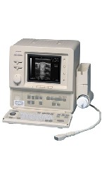 From ALOKA Co., Ltd.;
'A High-Quality, Portable Veterinary Ultrasound System. The SSD-500V is a rugged, portable convex sector/linear scanner that offers versatility, superb image quality and reliable performance in a compact design. This 22 lb system has Electronic dynamic focus and 256 shades of gray that produces high-quality images. The SSD-500V includes comprehensive measurement and analysis packages. Measurements include distance/depth, area, circumference, velocity, heart rate, ratio and angle. The system also included obstetrical analysis functions that can calculate estimated gestational age and expected date of confinement. In addition, customized obstetrical tables can be easily created. The SSD-500V software package also includes left ventricular function software for M-mode Cardiac evaluation. The 500V provides excellent clinical versatility − for use in both small and large animal applications − using a wide range of optional convex and linear array transducers.' •  [This entry is marked for removal.]
GE Medical Systems and Amersham announced in April 2004 the completion of a share exchange acquisition of Amersham Health by GE. The result of this acquisition is the new GE Healthcare, based in the UK, totally owned by General Electric (GE). The British company was a producer of contrast-imaging agents used to enhance image quality in X-ray, magnetic resonance imaging, and ultrasound procedures. It was also a leading producer of radiopharmaceuticals used in nuclear medicine imaging. Amersham Health was the firm's imaging, diagnostics, and therapeutics segment. Amersham was involved in biotechnology research through its Amersham Biosciences unit, which makes scanners, sequencers, microarrays, industrial separations, and other research supplies. • 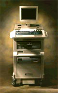 From SIUI Inc.;
'The Apogee 800Plus ultrasound system offers a new level of high quality in image. With broad clinical application, the system delivers ease of use for cardiac and vascular exams.' 'Due to the exceptional image quality, the sensitivity of color Doppler and many other advanced features, the Apogee 800Plus offers complete diagnosis capability for your entire patient population, including abdominal, obstetrics, gynecology, adult cardiology, pediatric cardiology, peripheral vascular, small parts, breast, prostate, transcranial, etc.'
Device Information and Specification
APPLICATIONS
Linear, curved, phased and symmetric phased array
2D, M-mode, Pulsed wave Doppler, Color Power Angio Imaging, Color 2D, Color M-mode, High PRF Doppler
H*W*D m
1.46 * 0.69 * 0.87
WEIGHT
140 kg (without peripherals)
POWER REQUIREMENT
115Vac, (V~), @ 50 to 60 Hz, 1.2kVA
230Vac, (V~), @ 50 to 60 Hz, 1.2kVA POWER CONSUMPTION
800 Watts (with optional OEM'S: 1200 Watts max)
•
An image artifact is any image attribute, which is not present in the original imaged object. An image artifact is sometime the result of an improper operation of the imager, and in other times a consequence of natural processes or properties of the human body. Artifacts in diagnostic ultrasound are a reflection or an echo, which appears on the display and represents the real anatomical structure not correctly. An artifact can be a false, multiple or misleading information introduced by the imaging system or by interaction of ultrasound with the adjacent tissue. Artifacts in ultrasound can be classified as to their source like e.g.:
•
physiologic (motion, different sound velocities, acoustical impedances of tissue);
•
hardware (dimension of the ultrasound beam and the transducer array);
•
Image artifacts can occur in each medical ultrasound. Then an interpretation of the image is complicated and can eliminate the structural information of objects looking for. See also Ultrasound Imaging Procedures. •
Beam steering is a method of steering the main lobe of a transducer to a certain direction. Array transducers have the ability to be steered as well as focused. Like focusing, the beam is directed by sequentially stimulation of each element. This feature creates the sector scan by rapidly steering the beam from left to right to give the two dimensional cross sectional image.
 Further Reading: Basics:
News & More:
Result Pages : | Share This Page     Look Ups |
 (.pdf)
Medical-Ultrasound-Imaging.com
former US-TIP.com
Member of SoftWays' Medical Imaging Group - MR-TIP • Radiology TIP • Medical-Ultrasound-Imaging
Copyright © 2008 - 2025 SoftWays. All rights reserved.
Terms of Use | Privacy Policy | Advertise With Us
former US-TIP.com
Member of SoftWays' Medical Imaging Group - MR-TIP • Radiology TIP • Medical-Ultrasound-Imaging
Copyright © 2008 - 2025 SoftWays. All rights reserved.
Terms of Use | Privacy Policy | Advertise With Us
[last update: 2025-04-05 02:38:00]