Medical Ultrasound Imaging
Monday, 29 April 2024
Info Sheets Out- side     | 'Averaging' Searchterm 'Averaging' found in 9 articles 1 term [ • ] - 8 definitions [• ] Result Pages : •
Frame averaging is a form of lowpass filtering. With frame averaging temporal variation are smoothed by addition of consecutive frames in a real-time mode.
•
The dimension of the ultrasound beam and the transducer array are the origin of the beam width artifact or volume averaging artifact. When the ultrasound beam is wider than the diameter of the lesion being scanned, normal tissues which lie immediately adjacent to the lesion arc included within the beam width, and their echotexture is averaged in with that of the lesion. Thus, what appears to be the echogenicity of the lesion is really that of the lesion plus the averaged normal tissues. Because of volume averaging, cystic lesions may falsely appear to be solid, and some subtle solid lesions may become impossible to distinguish from surrounding normal tissue and, therefore, not identified at all. See also Ultrasound Picture and Vector Array Transducer. •
The wider the ultrasound beam, the more severe the problem with volume averaging and the beam-width artifact, to avoid this, the ultrasound beam can be shaped with lenses.
Different possibilities to focus the beam:
•
Mechanical focusing is performed by placing an acoustic lens on the surface of the transducer or using a transducer with a concave face.
•
Electronic focusing uses multiple phased array (annular or linear) elements, sequentially fired to focus the beam.
Conventional multi-element transducers are electronically focused in order to minimize beam width. This transducer type can be focused electronically only along the long axis of the probe where there are multiple elements, along the short axis (elevation axis) are conventional transducers only one element wide. Electronic focusing in any axis requires multiple transducer elements arrayed along that axis. Short axis focusing of conventional multi-element transducers requires an acoustic lens which has a fixed focal length. For operation at frequencies at or even above 10 MHz, quantization noise reduces contrast resolution. Digital beamforming gives better control over time delay quantization errors. In digital beamformers the delay accuracy is improved, thus allowing higher frequency operation. In analog beamformers, delay accuracy is in the order of 20 ns. Phased beamformers are suitable to handle linear phased arrays and are used for sector formats such as required in cardiography to improve image quality. Beamforming in ultrasound instruments for medical imaging uses analog delay lines. The signal from each individual element is delayed in order to steer the beam in the desired direction and focuses the beam. The receive beamformer tracks the depth and focuses the receive beam as the depth increases for each transmitted pulse. The receive aperture increase with depth. The lateral resolution is constant with depth, and decreases the sensitivity to aberrations in the imaged tissue. A requirement for dynamic control of the used elements is given. Since often a weighting function (apodization) is used for side lobe reduction, the element weights also have to be dynamically updated with depth. See also Huygens Principle. •
Compound B-mode imaging takes different forms and refers to different methods of creating the ultrasound image. Real-time compound ultrasound improves the image quality of B-mode scanning by combining ultrasound information obtained from multiple angles. The used averaging process of compound B-mode reduces artifacts and improves the representation of true image data. B-mode images and Doppler mode images (see also Duplex) can be compounded on the display to improve the visualization of the anatomical relationships between vessels and the surrounding tissues. Further Reading: News & More:
•  Hitachi Medical Systems America Inc.;
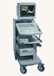
The EUB-525 is a mid-size ultrasound system with a full feature set. Capable of B-mode, M-mode, Doppler, color flow and color angiography it performs a variety of tasks in a simple and straightforward fashion. This system produces high quality images with excellent clarity and detail. Device Information and Specification
CLINICAL APPLICATION
CONFIGURATION
Compact, mobile system
Triple frequency
Linear, convex, radial, miniradial/miniprobe, bi-plane, echoendoscope longitudinal, echoendoscope radial
PROBE PORTS
Three
B/M-mode/simultaneous B/M-mode, CFM/CFA (color flow angiography)
IMAGING OPTIONS
Simultaneous imaging with biplane probes
IMAGING ENHANCEMENTS
Half-pitch scanning, dual scan line density, scan correlation and averaging, selectable receive filters
Result Pages : | Share This Page Look Ups |
Medical-Ultrasound-Imaging.com
former US-TIP.com
Member of SoftWays' Medical Imaging Group - MR-TIP • Radiology TIP • Medical-Ultrasound-Imaging
Copyright © 2008 - 2024 SoftWays. All rights reserved.
Terms of Use | Privacy Policy | Advertise With Us
former US-TIP.com
Member of SoftWays' Medical Imaging Group - MR-TIP • Radiology TIP • Medical-Ultrasound-Imaging
Copyright © 2008 - 2024 SoftWays. All rights reserved.
Terms of Use | Privacy Policy | Advertise With Us
[last update: 2023-11-06 01:42:00]