Medical Ultrasound Imaging
Sunday, 19 May 2024
Info Sheets Out- side     | 'Color Map' p4 Searchterm 'Color Map' found in 19 articles 1 term [ • ] - 4 definitions [• ] - 14 booleans [• ]Result Pages : •
Variance is the modification of Doppler frequencies within each pixel during a pulse packet with color Doppler flow imaging. Variance is mapped to the color green and is used to detect turbulence. At low variance (laminar flow) pixels within each range gate are encoded in red or blue according to flow direction. At high variance (turbulent flow) a percentage of pixels within each range gate are encoded with green according to the degree of spectral broadening.
•  From ESAOTE S.p.A.;
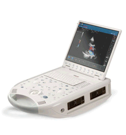
'The MyLab™30CV ultrasound system is an evolutionary step in ultrasound technology. Weighing less than 20 pounds, it is the first compact ultrasound system to deliver premium console performance. And with mobile, portable or stationary configurations, MyLab30CV can adapt to any clinical environment.'
Device Information and Specification
APPLICATIONS
Abdominal, breast, cardiac, OB/GYN, pediatric, pediatric cardiology, small parts, transcranial, vascular
CONFIGURATION
Portable
Linear: 4-10 MHz, convex: 2-5 MHz, phased: 1.6-10 MHz, micro convex: 5-7.5 MHz, endocavity: 5-7.5 MHz, pencil: 2 + 5 MHz
2-D, M-mode, duplex, triplex, color Doppler, pulsed wave Doppler, tissue velocity mapping (TVM), tissue enhancement imaging (TEI™), contrast harmonic imaging, stress echo, tissue velocity mapping for LV motion analysis (TVM), contrast tuned imaging for contrast media procedures (CnTI™), Qontrast™ for myocardium parameters quantification
STORAGE, CONNECTIVITY, OS
Digital patient archive/management, integrated CD/RW, RJ 45 and USB ports, Windows
H*W*D m (inch.)
0.16 * 0.36 * 0.50 (6.2 x 14 x 19.3)
WEIGHT
Less than 11 kg (20 lbs.)
•  From Kontron Medical SAS;
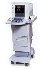
'The Sigma 330 is a versatile, digital, mobile Ultrasound System, upgradeable from 2D to Doppler, Color Flow Mapping and 3D. The Sigma 330 displays excellent image quality and superb Doppler and CFM sensitivity, employing ATEC™ (Advanced Tissue Echo Cancellation), a revolutionary technology developed by the Kontron Medical R&D Center, which removes tissue artifacts ('ghosting').' Specifications for this system will be available soon. •
Ultrasound elastography is a specialized imaging technique that provides information about tissue elasticity or stiffness. It is used to assess the mechanical properties of tissues, helping to differentiate between normal and abnormal tissue conditions.
The basic principle behind ultrasound elastography involves the application of mechanical stress to the tissue and measuring its resulting deformation. This is typically achieved by using either external compression or shear waves generated by the ultrasound transducer. There are two main types of ultrasound elastography: •
Strain Elastography: In strain elastography, the tissue is mechanically compressed using the ultrasound transducer, causing deformation. The transducer then captures images before and after compression, and the software analyzes the displacement or strain between these images. Softer tissues tend to deform more than stiffer tissues, and this information is used to generate a color-coded map or elastogram, where softer areas appear in different colors compared to stiffer regions.
•
Shear Wave Elastography: Shear wave elastography involves the generation of shear waves within the tissue using focused ultrasound beams. These shear waves propagate through the tissue, and their velocity is measured using the ultrasound transducer. The speed of shear wave propagation is directly related to tissue stiffness: stiffer tissues transmit shear waves faster than softer tissues. By calculating the shear wave velocity, an elastogram is generated, providing a quantitative assessment of tissue stiffness.
Both strain elastography and shear wave elastography offer valuable insights into tissue characteristics and can assist in the diagnosis and characterization of various conditions. In clinical practice, ultrasound elastography is particularly useful for evaluating liver fibrosis, breast lesions, thyroid nodules, prostate abnormalities, and musculoskeletal conditions. By providing additional information about tissue stiffness, ultrasound elastography enhances the diagnostic capabilities of traditional ultrasound imaging. It allows for non-invasive assessment, improves the accuracy of tissue characterization, and aids in treatment planning and monitoring of various medical conditions. See also Ultrasound Accessories and Supplies, Sonographer and Ultrasound Technology. Result Pages : | Share This Page Look Ups |
Medical-Ultrasound-Imaging.com
former US-TIP.com
Member of SoftWays' Medical Imaging Group - MR-TIP • Radiology TIP • Medical-Ultrasound-Imaging
Copyright © 2008 - 2024 SoftWays. All rights reserved.
Terms of Use | Privacy Policy | Advertise With Us
former US-TIP.com
Member of SoftWays' Medical Imaging Group - MR-TIP • Radiology TIP • Medical-Ultrasound-Imaging
Copyright © 2008 - 2024 SoftWays. All rights reserved.
Terms of Use | Privacy Policy | Advertise With Us
[last update: 2023-11-06 01:42:00]