Medical Ultrasound Imaging
Sunday, 19 May 2024
Info Sheets Out- side     | 'Digital' p3 Searchterm 'Digital' found in 63 articles 4 terms [ • ] - 59 definitions [• ] Result Pages : • 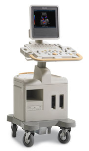 From Philips Medical Systems;
Introduced in June 2005, 'one of the less expensive and more dedicated' ultrasound systems. •
(PACS) A system used to communicate and archive medical imaging data, mostly images and associated textural data generated in a radiology department, and disseminated throughout the hospital. A PACS is usually based on the DICOM (Digital Imaging and Communications in Medicine) standard. The main components in the PACS are: acquisition devices where the images are acquired;
•
short and longer term archives for storage of digital and textural data;
•
a database and database management;
•
diagnostic and review workstations;
•
software to run the system;
•
a communication network linking the system components;
•
interfaces with other networks (hospital and radiological information systems).
Acquisition devices, which acquire their data in direct digital format, like a MRI system, are most easily integrated into a PACS. Short term archives need to have rapid access, such as provided by a RAID (redundant array of independent disks), whereas long term archives need not have such rapid access and can be consigned, e.g. to optical disks or a magnetic. High speed networks are necessary for rapid transmission of imaging data from the short term archive to the diagnostic workstations. Optical fibre, ATM (asynchronous transfer mode), fast or switched Ethernet, are examples of high speed transmission networks, whereas demographic textural data may be transmitted along conventional Ethernet. Sophisticated software is a major element in any hospital-wide PACS. The software concepts include: preloading or prefetching of historical images pertinent to current examinations, worklists and folders to subdivide the vast mass of data acquired in a PACS in a form, which is easy and practical to access, default display protocols whereby images are automatically displayed on workstation monitors in a prearranged clinically logical order and format, and protocols radiologists can rapidly report worklists of undictated examinations, using a minimum of computer manipulation. • 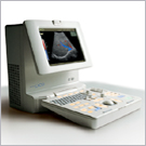 From Medison Co.,Ltd.;
'Full-featured Digital Imaging On Your Desktop The SONOACE PICO is a full-featured digital color ultrasound system with virtually all the imaging capabilities you would expect from a cart-based system with the added benefit of being fully portable. Built on a reliable Linux PC platform, the SONOACE PICO boasts all- digital beamforming and signal processing for best-in-class image resolution enhanced by a long list of advanced diagnostic tools such as harmonic imaging, color and power Doppler, and freehand 3D imaging.' • 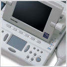 From Medison Co.,Ltd.;
'Putting the Power of Portable Ultrasound in Veterinarians' Hands Personal Veterinary Digital Ultrasound Yes, SONOVET2000 is good news for our four-legged friends, veterinarians and animal husbandry professionals. Portable and wearable, it puts the power of digital ultrasound scanning in your hands for non-invasive diagnosis anywhere, anytime. With it's on-site capabilities, SONOVET2000 raises the bar for animal health care quality and your working efficiency. And it comes with the superior imaging power of Medison's all-digital beamforming technology. Enjoy a higher productivity and efficiency at a surprisingly competitive price.' •
Ultrasound machines, widely used in medical imaging, are essential tools in the field of diagnostic ultrasound. These devices utilize high-frequency sound waves to create real-time images of internal body structures. Ultrasound machines consist of several key components that work together to generate diagnostic images.
These include:
•
The transducer is a handheld device that emits and receives sound waves. It converts electrical energy into sound waves and captures the returning echoes to create images.
•
The control panel houses the interface where the sonographer adjusts imaging parameters such as depth, frequency, and gain. It allows for customization of imaging settings based on the clinical requirements. The transducer pulse controls change the amplitude, frequency and duration of the pulses emitted from the transducer probe.
•
The central processing unit (CPU) serves as the brain of the ultrasound machine, processing the acquired data and transforming it into images. It handles complex calculations, image optimization, data storage and contains the electrical power supplies for itself and the transducer probe.
•
The display monitor (oscilloscope, tablet, computer monitor, etc.) showcases the real-time ultrasound images produced by the machine. It provides visual feedback to the sonographer, aiding in the interpretation and analysis of anatomical structures. Handheld ultrasound devices and mobile ultrasound probes can be connected wirelessly to a smartphone or tablet via Bluetooth or WiFi. These end device serves then as the ultrasound monitor.
•
Data input and measurements are done with the keyboard cursor (trackball). Ultrasound devices used for handheld point of care ultrasound (HPOCUS) are operated via the touch screen of the control panel.
•
Images are captured, reviewed, stored and transmitted digitally, using a standard format for digital imaging and communications in medicine (DICOM). Disk storage devices (FDD, HDD, CD, DVD) are outdated, but may be used in older machines to store the acquired images if no picture archiving and communication system (PACS) connection is possible.
•
The displayed ultrasound pictures are usually digitally stored in a PACS. The images from portable ultrasound machines can be stored and conveniently managed on the end device itself, the inserted memory card or in the cloud. With a QR scanner, the images can be accessed via the Internet in the cloud. Often there is also the possibility to get a picture of a baby sonography as a printout.
B-mode machines represent the vast majority of machines used in echocardiology, obstetrical scans, abdominal scans, gynecological scans, etc. B-mode ultrasound machines usually produce the sector (or pie segment-shaped) scans. These ultrasound scans require either a mechanical scanner transducer (the transducer moves to produce the sector scan), or a linear array transducer operated as a phased array. Ultrasound machines come in different types, each catering to specific clinical needs. The two primary types are stationary and portable ultrasound machines: •
Stationary units are typically larger in size and are installed in dedicated imaging rooms. These machines offer advanced imaging capabilities and a wide range of specialized features. They are commonly found in hospitals, clinics, and university medical centers where comprehensive imaging services are provided.
•
Portable units (see Portable Ultrasound Machine), as the name suggests, are compact and lightweight, designed for on-the-go imaging. These machines are highly versatile and offer excellent mobility, allowing healthcare professionals to bring the ultrasound system directly to the patient's bedside. Portable ultrasound machines are particularly useful in emergency settings, rural healthcare facilities, and point-of-care applications.
See also Handheld Ultrasound, Ultrasound System Performance, Equipment Preparation, Coaxial Cable, and Microbubble Scanner Modification, Environmental Protection and Ultrasound Accessories and Supplies. Further Reading: Basics: News & More:
Result Pages : | Share This Page Look Ups |
Medical-Ultrasound-Imaging.com
former US-TIP.com
Member of SoftWays' Medical Imaging Group - MR-TIP • Radiology TIP • Medical-Ultrasound-Imaging
Copyright © 2008 - 2024 SoftWays. All rights reserved.
Terms of Use | Privacy Policy | Advertise With Us
former US-TIP.com
Member of SoftWays' Medical Imaging Group - MR-TIP • Radiology TIP • Medical-Ultrasound-Imaging
Copyright © 2008 - 2024 SoftWays. All rights reserved.
Terms of Use | Privacy Policy | Advertise With Us
[last update: 2023-11-06 01:42:00]