Medical Ultrasound Imaging
Sunday, 19 May 2024
Info Sheets Out- side     | 'Enhancement' p2 Searchterm 'Enhancement' found in 28 articles 2 terms [ • ] - 26 definitions [• ] Result Pages : •
(UCA / USCA) Ultrasonography is the most commonly performed diagnostic imaging procedure. The introduction of sonographic contrast media into routine practice modifies the use of ultrasound in a variety of clinical applications. USCAs consist of microbubbles filled with air or gases and can be classified according to their pharmacokinetics. Among the blood pool agents, transpulmonary ultrasound contrast agents offer higher diagnostic potential compared to agents that cannot pass the pulmonary capillary bed after a peripheral intravenous injection. In addition to their vascular phase, some USCAs can exhibit a tissue- or organ-specific phase. The sonogram image quality is improved either by decreasing the reflectivity of the undesired interfaces or by increasing the backscattered echoes from the desired regions. Different types of ultrasound contrast agents: Ultrasound contrast agents act as echo-enhancers, because of the high different acoustic impedance at the interface between gas and blood. The enhanced echo intensity is proportional to the change in acoustical impedance as the sound beam crosses from the blood to the gas in the bubbles. The ideal qualities of an ultrasound contrast agent:
•
high echogenicity;
•
low attenuation;
•
low blood solubility;
•
low diffusivity;
•
ability to pass through the pulmonary capillary bed;
•
lack of biological effects with repeat doses.
A typical ultrasound contrast agent consists of a thin flexible or rigid shell composed of albumin, lipid, or polymer confining a gas such as nitrogen, or a perfluorocarbon. The choice of the microbubble shell and gas has an important influence on the properties of the agent. Current generations of microbubbles have a diameter from 1 μm to 5 μm. The success of these agents is mostly dependent on the small size and on the stability of their shell, which allows passage of the microbubbles through the pulmonary circulation. Microbubbles must be made smaller than the diameter of capillaries or they would embolize and be ineffective and perhaps even dangerous. The reflectivity of these microbubbles is proportional to the fourth power of a particle diameter but also directly proportional to the concentration of the contrast agent particles themselves. Ultrasound contrast agents produce unique acoustic signatures that allow to separate their signal from tissue echoes and to depict whether they are moving or stationary. This enables the detection of capillary flow and of targeted microbubbles that are retained in tissues such as normal liver. The new generation of contrast media is characterized by prolonged persistence in the vascular bed which provides consistent enhancement of the arterial Doppler signal. Contrast agents make it also possible to perform dynamic and perfusion studies. Targeted contrast imaging agents are for example taken up by the phagocytic cell systems and thus have liver/spleen specific effects. See also Ultrasound Contrast Agent Safety, Adverse Reaction, Tissue-Specific Ultrasound Contrast Agent, and Bubble Specific Imaging. Further Reading: Basics:
News & More:
•
Vascular ultrasound contrast agents are gas microbubbles with a diameter less than 10 μm (2 to 5 μm on average for most of the newer agents) to pass through the lung capillaries and enter into the systemic circulation. Air bubbles in that size persist in solution for only a short time; too short for systemic vascular use in medical ultrasound imaging. So the gas bubbles have to be stabilized to persist long enough and survive pressure changes in the heart. Most vascular contrast media are stabilized against dissolution and coalescence by the presence of additional materials at the gas-liquid interface. In some cases, this material is an elastic solid shell that enhances stability by supporting a strain to counter the effect of surface tension. In other cases, the material is a surfactant, or a combination of two or more surfactants. Typically the effective duration of vascular enhancement is a few minutes, after which the microbubbles dissipate. This rather short duration of vascular enhancement makes it easy to perform repeated dynamic studies. Intravenous vascular contrast agents will be used in imaging malignant tumors in the liver, kidney, ovary, pancreas, prostate, and breast. Tumor neovascularization can be a marker for angiogenesis, and Doppler signals from small tumor vessels may be detectable after contrast injection. Contrast agents are useful for evaluating vessels in a variety of organs, including those involved in renal, hepatic, and pancreatic transplants. If an area of ischemia or a stenosis is detected after contrast administration, the use of other more expensive imaging modalities, including CT and MRI, can often be avoided. See also Acoustically Active Lipospheres. •
(CAP) Contrast agent particles are filled with gas and coated by a shell. The reflectivity of a particle increases with the diameter. The used particles are too large to cross the endothelium, so that there is no interstitial phase of enhancement. They are essentially markers for the blood pool and their distribution is similar to those of tagged red cells. In addition, any body cavity that can be accessed can be injected with vascular contrast.
•
(CEUS) Contrast agents increase the reflection of ultrasonic energy, improve the signal to noise ratio and caused by that the detection of abnormal microvascular and macrovascular disorders. Contrast enhanced ultrasound is used in abdominal ultrasound (liver sonography) as well as in cerebrovascular examinations e.g., for an accurate grading of carotid stenosis. The used contrast agents are safe and well tolerated. The quality of the enhancement depends on:
•
the concentration of the contrast agent;
•
the type of injection, flow rate;
•
the patient characteristics;
•
the microbubble quality and properties of the filling gas and the shell.
The additional use of ultrasound contrast agents (USCAs) may overcome typical limitations like poor contrast of B-mode imaging or limited sensitivity of Doppler techniques. The development of new ultrasound applications (e.g., blood flow imaging, perfusion quantification) depends also from the development of pulse sequences for bubble specific imaging. In addition, contrast enhanced ultrasound improves the monitoring of ultrasound guided interventions like RF thermal ablation. See also Contrast Enhanced Doppler Imaging, Contrast Harmonic Imaging, Contrast Imaging Techniques and Contrast Pulse Sequencing. Further Reading: News & More:
• 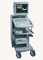 Hitachi Medical Systems America Inc.;
The EUB-525 is a mid-size ultrasound system with a full feature set. Capable of B-mode, M-mode, Doppler, color flow and color angiography it performs a variety of tasks in a simple and straightforward fashion. This system produces high quality images with excellent clarity and detail. Device Information and Specification
CLINICAL APPLICATION
CONFIGURATION
Compact, mobile system
Triple frequency
Linear, convex, radial, miniradial/miniprobe, bi-plane, echoendoscope longitudinal, echoendoscope radial
PROBE PORTS
Three
B/M-mode/simultaneous B/M-mode, CFM/CFA (color flow angiography)
IMAGING OPTIONS
Simultaneous imaging with biplane probes
IMAGING ENHANCEMENTS
Half-pitch scanning, dual scan line density, scan correlation and averaging, selectable receive filters
Result Pages : | Share This Page Look Ups |
Medical-Ultrasound-Imaging.com
former US-TIP.com
Member of SoftWays' Medical Imaging Group - MR-TIP • Radiology TIP • Medical-Ultrasound-Imaging
Copyright © 2008 - 2024 SoftWays. All rights reserved.
Terms of Use | Privacy Policy | Advertise With Us
former US-TIP.com
Member of SoftWays' Medical Imaging Group - MR-TIP • Radiology TIP • Medical-Ultrasound-Imaging
Copyright © 2008 - 2024 SoftWays. All rights reserved.
Terms of Use | Privacy Policy | Advertise With Us
[last update: 2023-11-06 01:42:00]