Medical Ultrasound Imaging
Sunday, 19 May 2024
Info Sheets Out- side     | 'Gain' p2 Searchterm 'Gain' found in 19 articles 2 terms [ • ] - 17 definitions [• ] Result Pages : •
B-scan comined with D-scan (D=Depth) is used to avoid image inhomogeneity. Different transmitter signals for each depth are applied and prefiltered pseudoinversely according to the transfer properties of the covering tissue. Pulse compression techniques with nonlinearly frequency modulated signals are used to gain the required energy for inverse filtering. D-scan is a modified C-scan used in nondestructive testing with the display of amplitudes. In the 2D graphical presentation, time of flight values are displayed in the top view on a test surface. See also A-Scan, B-Scan and C-Scan. • 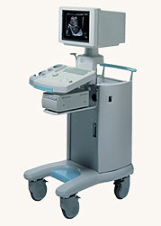 From Hitachi Medical Systems America Inc.;
The EUB-500 is similar to the EUB-405 PLUS. Added features are the 12-inch monitor, the second probe port, cine memory, gain slide control/multifunction trackball.
Device Information and Specification
APPLICATIONS
General purpose, OB, urological
CONFIGURATION
Compact portable system
Dual frequency
Linear, convex, radial, bi-plane, echoendoscope longitudinal
PROBE PORTS
Two
OPTIONAL PACKAGE
Multi-memory, digital image storage and transfer via flashcard, cart
WEIGHT
Less than 14 kg
•
A liver sonography is a diagnostic tool to image the liver and adjoining upper abdominal organs such as the gallbladder, spleen, and pancreas. Deeper structures such as liver and pancreas are imaged at a lower frequency 1-6 MHz with lower axial and lateral resolution but greater penetration. The diagnostic capabilities in this area can be limited by gas in the bowel scattering the sound waves. The application of microbubbles may be useful for detection of liver lesions and for lesion characterization. Some microbubbles have a liver-specific post vascular phase where they appear to be taken up by the reticuloendothelial system (RES). Dynamic contrast enhanced scans in a similar way as with CT or MRI can be used to studying the arterial, venous and tissue phase. After a bolus injection, early vascular enhancement is seen at around 30sec in arterialized lesions (e.g., hepatocellular carcinomas (HCC), focal nodular hyperplasia (FNH)). Later enhancement is typical of hemangiomas with gradually filling towards the center. In the late phase at around 90sec, HCCs appear as defects against the liver background. Most metastases are relatively hypovascular and so do not show much enhancement and are seen as signal voids in the different phases. Either with an intermittent imaging technique or by continuous scanning in a nondestructive, low power mode, characteristic time patterns can be used to differentiate lesions. See also Medical Imaging, B-Mode, High Intensity Focused Ultrasound, Ultrasound Safety and Contrast Medium. Further Reading: Basics:
News & More:
•
A microbubble shell, designed to reduce diffusion into the blood, can be stiff (e.g., denatured albumin) or more flexible (phospholipid), varying in thickness from 10-200 nm. The shell stabilizes against dissolution and coalescence with additional materials at the gas-liquid interface. This material can be an elastic solid shell that enhances stability by supporting a strain to counter the effect of surface tension. Also a surfactant, or a combination of two or more, improves the stability by a high reduction of the surface tension at the interface. Current ultrasound contrast agents are micron-sized bubbles with a stabilizing shell. Further Reading: Basics: News & More:
•
The mirror artifact is similar to the reverberation artifact. Mirror image artifacts (mirroring) can occur if the acoustical impedances of the tissue is too much different and the ultrasound is reflected multiple times on tissue layers.
The echo detected does not come from the shortest sound path, the sound is reflected off an angle to another interface so that like a real mirror, the artifact shows up as the virtual object. An empyema or lung abscess can be simulated by a mirror image artifact of a hepatic cyst. This liver lesion can appear like a lesion within the lung because the wave is reflected off the diaphragm back into the liver. The angle of reflection is equal to the angle of incidence. The sound pulse hits the interfaces within the liver lesion and is reflected back to the diaphragm once again with an angle of reflection equal to the angle of incidence and then back to the transducer. Also by a pelvic ultrasound scan the sound can be reflected off the rectal air at an angle so that the deep wall of an artifactual cyst represents the mirror image of the inferior and anterior walls of the bladder. Mirror image artifacts can cause other strange appearances such as invasion of a transitional cell carcinoma through the bladder wall. Also called Cross Talk. Further Reading: News & More:
Result Pages : | Share This Page Look Ups |
Medical-Ultrasound-Imaging.com
former US-TIP.com
Member of SoftWays' Medical Imaging Group - MR-TIP • Radiology TIP • Medical-Ultrasound-Imaging
Copyright © 2008 - 2024 SoftWays. All rights reserved.
Terms of Use | Privacy Policy | Advertise With Us
former US-TIP.com
Member of SoftWays' Medical Imaging Group - MR-TIP • Radiology TIP • Medical-Ultrasound-Imaging
Copyright © 2008 - 2024 SoftWays. All rights reserved.
Terms of Use | Privacy Policy | Advertise With Us
[last update: 2023-11-06 01:42:00]