Medical Ultrasound Imaging
Wednesday, 8 May 2024
Info Sheets Out- side     | 'Power Mode' Searchterm 'Power Mode' found in 21 articles 1 term [ • ] - 2 definitions [• ] - 18 booleans [• ]Result Pages : • Power Mode
In power mode the amplitude (power) of color Doppler signals is displayed, regardless of the velocity. Power does not have negative values and is independent of sampling frequency. An aliasing artifact does not occur in power mode images. Caused by plotting the quantity enhanced by echo contrast agents in a power map, power mode is often used in contrast Doppler ultrasound examinations. Also known as energy mode. • View NEWS results for 'Power Mode' (3). •
A liver sonography is a diagnostic tool to image the liver and adjoining upper abdominal organs such as the gallbladder, spleen, and pancreas. Deeper structures such as liver and pancreas are imaged at a lower frequency 1-6 MHz with lower axial and lateral resolution but greater penetration. The diagnostic capabilities in this area can be limited by gas in the bowel scattering the sound waves. The application of microbubbles may be useful for detection of liver lesions and for lesion characterization. Some microbubbles have a liver-specific post vascular phase where they appear to be taken up by the reticuloendothelial system (RES). Dynamic contrast enhanced scans in a similar way as with CT or MRI can be used to studying the arterial, venous and tissue phase. After a bolus injection, early vascular enhancement is seen at around 30sec in arterialized lesions (e.g., hepatocellular carcinomas (HCC), focal nodular hyperplasia (FNH)). Later enhancement is typical of hemangiomas with gradually filling towards the center. In the late phase at around 90sec, HCCs appear as defects against the liver background. Most metastases are relatively hypovascular and so do not show much enhancement and are seen as signal voids in the different phases. Either with an intermittent imaging technique or by continuous scanning in a nondestructive, low power mode, characteristic time patterns can be used to differentiate lesions. See also Medical Imaging, B-Mode, High Intensity Focused Ultrasound, Ultrasound Safety and Contrast Medium. Further Reading: Basics:
News & More:
• • 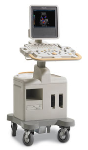 From Philips Medical Systems;
Introduced in June 2005, 'one of the less expensive and more dedicated' ultrasound systems.
Device Information and Specification
CONFIGURATION
LCD monitor
Broadband, convex, linear,
digital beamformer and focal tuning IMAGING OPTIONS
OPTIONAL PACKAGE
DICOM, etc.
STORAGE, CONNECTIVITY, OS
HDD, CD, USB, optionalMOD and DICOM 3.0
DATA PROCESSING
256-digitally processed channels
H*W*D inch.
58 * 20 * 32
WEIGHT
135 lbs.
• 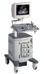 From ALOKA Co., Ltd.;
'A Platform for Digital, Pure-Beam Imaging The high-performance, ALOKA ProSound SSD-3500 utilizes advanced ProSound technologies including: Fully digital beam former A wide dynamic range, 12-bit A/D converter Multi beam processing. The SSD-3500 also helps you achieve more efficient examinations. Its ergonomic, user-friendly design enables you to customize the system according to your specific application needs.'
Device Information and Specification
APPLICATIONS
CONFIGURATION
Compact, portable, dual dynamic display
Color Flow, Power Flow, Spectral Doppler, Real-time Free Angular M-Mode, Tissue Harmonic Imaging, Quint Frequency Imaging, Pure Harmonic Detection
STORAGE, CONNECTIVITY, OS
Data Management Subsystem (iDMS), DICOM-Worklist
DATA PROCESSING
12-bit analog to digital converter
Result Pages : | Share This Page Look Ups |
Medical-Ultrasound-Imaging.com
former US-TIP.com
Member of SoftWays' Medical Imaging Group - MR-TIP • Radiology TIP • Medical-Ultrasound-Imaging
Copyright © 2008 - 2024 SoftWays. All rights reserved.
Terms of Use | Privacy Policy | Advertise With Us
former US-TIP.com
Member of SoftWays' Medical Imaging Group - MR-TIP • Radiology TIP • Medical-Ultrasound-Imaging
Copyright © 2008 - 2024 SoftWays. All rights reserved.
Terms of Use | Privacy Policy | Advertise With Us
[last update: 2023-11-06 01:42:00]