Medical Ultrasound Imaging
Wednesday, 15 May 2024
Info Sheets Out- side     | 'Hertz' p2 Searchterm 'Hertz' found in 12 articles 1 term [ • ] - 11 definitions [• ] Result Pages : •
A logarithmic measure of sound loudness closely related to the decibel. The unit decibel is used for objective measurements, that means, they measure the actual pressure of the sound waves as recorded using a microphone. The unit phon is used for subjective measurements, which means, measurements made using the ears of a human listener. A sound has the loudness 'p' phon if it seems to the listener to be equal in loudness to the sound of a pure tone of the frequency 1 kilohertz and strength 'p' decibel. A measurement in phons will be similar to a measurement in decibel, but not identical, since the perceived loudness of a sound depends on the distribution of frequencies in the sound as well as the pressure of the sound waves. In the U.S., sound loudness is frequently measured in sones rather than phons: a sound of loudness x sones has loudness 10 log2 x + 40 phons. See also Acoustic Noise. • View NEWS results for 'Phon' (9). • 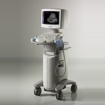 From Siemens Medical Systems;
'The SONOLINE G20™ ultrasound system quickly distances itself from the competition with next-generation all-digital system architecture that utilizes Siemens technology migration. Individual imaging parameters have been optimized for a wide variety of clinical applications and patient types. So you can realize a higher degree of diagnostic confidence. Without doubt.'
Device Information and Specification
CONFIGURATION
Compact, ultra-portable system
MultiHertz™ multiple frequency
PROBE TYPES
MicroCase™ transducer
IMAGING OPTIONS
Tissue Harmonic Imaging (THI)
IMAGING ENHANCEMENTS
TGO™ tissue grayscale optimization technology
STORAGE
DIMAQ-IP integrated workstation
DATA PROCESSING
Powerful processor for rapid transition times
•
Sonography [aka: ultrasonography] is a term that encompasses the entire process of performing ultrasound examinations and interpreting the obtained images. Sonography involves the skilled application of ultrasound technology by trained professionals known as sonographers or ultrasound technologists. These specialists operate the ultrasound equipment, manipulate the transducer, and acquire the necessary pictures for diagnostic imaging purposes. Sonography requires in-depth knowledge of anatomy, physiology, and pathology to accurately interpret the ultrasound images and provide valuable information to the treating physician. Sonography uses equipment that generates high frequency sound waves to produce images from muscles, soft tissues, fluid collections, and vascular structures of the human body. Obstetric sonography is commonly used during pregnancy. Sonography visualizes anatomy, function, and pathology of for example gallbladder, kidneys, pancreas, spleen, liver, uterus, ovaries, urinary bladder, eye, thyroid, breast, aorta, veins and arteries in the extremities, carotid arteries in the neck, as well as the heart. A typical medical ultrasound machine, usually a real-time scanner, operates in the frequency range of 2 to 13 megahertz. See also Musculoskeletal and Joint Ultrasound, Pediatric Ultrasound, Cerebrovascular Ultrasonography and Contrast Enhanced Ultrasound. Further Reading: Basics:
News & More:
•
Sound and ultrasound waves consist of a mechanical disturbance of a medium such as air. The disturbance passes through the medium at a fixed speed causing vibration. The rate at which the particles vibrate is the frequency, measured in cycles per second or Hertz (Hz). The pressure of sound is reported on a logarithmic scale called sound-pressure level, expressed in decibel (dB) referenced to the weakest audible 1 000 Hz sound pressure of 2*10-5 Pascal (20 mP). Sound level meters contain filters that simulate the ear's frequency response. The most commonly used filter provides what is called 'A' weighting, with the letter 'A' appended to the dB units, i.e. dBA. Sound becomes inaudible to the human ear above about 20 kHz and is then known as ultrasound. Diagnostic imaging uses much higher frequencies, in the order of MHz. See also Spatial Peak Intensity. Sound frequencies:
•
infrasound - 0 to 20 Hz;
•
audible sound - 20 Hz to 20 KHz;
•
ultrasound - greater than 20 KHz;
•
medical ultrasound - 2.5 MHz to 15 MHz.
Further Reading: Basics: •
Submicron ultrasound contrast agents are gas-filled, double-walled microspheres with a diameter smaller than 1 μm that rupture when exposed
to ultrasound energy at megahertz frequencies. These agents differ from traditional ultrasound contrast microbubbles in that the submicron bubbles may serve as extravascular agents. They are small enough to travel through the lymphatic system and to be extravasated from tumor neovasculature. The detection of these agents is limited by their hard shell, which requires high-pressure ultrasound insonation
for shell rupture and excitation of the gas bubble. After shell rupture, the gas diffuses rapidly from submicron sized agents. The optimal processing of each echo is important.
Result Pages : | Share This Page Look Ups |
Medical-Ultrasound-Imaging.com
former US-TIP.com
Member of SoftWays' Medical Imaging Group - MR-TIP • Radiology TIP • Medical-Ultrasound-Imaging
Copyright © 2008 - 2024 SoftWays. All rights reserved.
Terms of Use | Privacy Policy | Advertise With Us
former US-TIP.com
Member of SoftWays' Medical Imaging Group - MR-TIP • Radiology TIP • Medical-Ultrasound-Imaging
Copyright © 2008 - 2024 SoftWays. All rights reserved.
Terms of Use | Privacy Policy | Advertise With Us
[last update: 2023-11-06 01:42:00]