Medical Ultrasound Imaging
Sunday, 19 May 2024
Info Sheets Out- side     | 'Kidney Ultrasound' p2 Searchterm 'Kidney Ultrasound' found in 15 articles 1 term [ • ] - 3 definitions [• ] - 11 booleans [• ]Result Pages : •  From SIUI Inc.;
'Dedicated to ultrasound industry, Shantou Institute of Ultrasonic Instruments, Inc. (SIUI) has launched Apogee 3500, the Digital Color Doppler Ultrasound Imaging System. With latest imaging technologies, high-definition image quality and excellent practical functions, the Apogee 3500 offers optimal solutions for clinical ultrasonic examination.' 'The Apogee 3500 is available with many high-density, super broadband and multi-frequency probes, such as convex, micro-convex, linear, vaginal, rectal and phased array probes, which are widely applied for different clinical diagnoses, including abdomen (liver, kidney, gall-bladder, pancreas), gynecology (uterus, ovary), obstetrics (early pregnancy, basic OB, complete OB, multi gestation, fetal echo), cardiology (adult and pediatric cardiology), small parts (thyroid, galactophore, testicles, neonate), peripheral vascular and prostate.'
Device Information and Specification
CONFIGURATION
Normal system, color - gray scale(256)
Linear, convex and phased array
PROBES STANDARD
2.0 MHz ~ 12.0 MHz, broad band, tri-frequency
B-mode (B, 2B, 4B), M-mode, B/M-mode, real-time compound imaging, panoramic imaging, trapezoidal imaging (linear probes), spectrum Doppler (PWD and CWD), color Doppler flow imaging (CDFI), color power angio (CPA), tissue harmonic imaging (THI)
IMAGING OPTIONS
Real-time ZOOM, zoom rate and position selectable
OPTIONAL PACKAGE
H*W*D m
1.29 * 0.52 * 0.75
WEIGHT
110 kg
POWER REQUIREMENT
AC 220V/110V, 50Hz/60Hz
POWER CONSUMPTION
0.6 KVA
•  From SIUI Inc.;
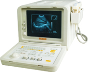
'SIUI's newly developed CTS-485 is a portable ultrasound system with advanced technology. With its powerful features, such as 256 grayscale, cineloop, RS232C interface and broadband high-density, multi-frequency probes, the system is designed for professional applications in cardiology, abdominal, small parts, liver, gallbladder, kidney, obstetrics, gynecology, peripheral vascular, etc. The CTS-485 has passed FDA clearance and CE Marking.'
Device Information and Specification
APPLICATIONS
CONFIGURATION
Portable, gray scale(256)
Linear and convex
PROBES STANDARD
1 * 2.5MHz ~ 5.0MHz multifrequency convex probe
2.5MHz to 10.0MHz broad band, trifrequency
IMAGING OPTIONS
Multi zoom rate and depth shift
OPTIONAL PACKAGE
H*W*D m
0.26 * 0.3 * 0.41
WEIGHT
11 kg (main unit)
POWER REQUIREMENT
AC 220V/110V, 50Hz/60Hz
POWER CONSUMPTION
0.1 KVA
•  From SIUI Inc.;
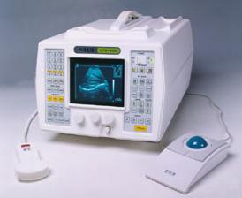
'The CTS-285 is designed for the diagnosis of liver, gallbladder, kidney, pancreas, thyroid, breast, uterus, bladder, ovary, etc. It is a portable versatile ultrasound scanner with convex and linear array scanning.'
Device Information and Specification
APPLICATIONS
See description above
CONFIGURATION
Portable, gray scale(256)
Linear and convex
PROBES STANDARD
1 * 2.5MHz ~ 5.0MHz trifrequency convex probe
2.5MHz to 10.0MHz, linear and convex, broad band, trifrequency
IMAGING OPTIONS
Multi zoom rate and depth shift
DATA PROCESSING
Pre-processing, correlation-processing, interpolation
POWER REQUIREMENT
AC 220V/110V, 50Hz/60Hz
POWER CONSUMPTION
0.06 KVA
•
Sonography [aka: ultrasonography] is a term that encompasses the entire process of performing ultrasound examinations and interpreting the obtained images. Sonography involves the skilled application of ultrasound technology by trained professionals known as sonographers or ultrasound technologists. These specialists operate the ultrasound equipment, manipulate the transducer, and acquire the necessary pictures for diagnostic imaging purposes. Sonography requires in-depth knowledge of anatomy, physiology, and pathology to accurately interpret the ultrasound images and provide valuable information to the treating physician. Sonography uses equipment that generates high frequency sound waves to produce images from muscles, soft tissues, fluid collections, and vascular structures of the human body. Obstetric sonography is commonly used during pregnancy. Sonography visualizes anatomy, function, and pathology of for example gallbladder, kidneys, pancreas, spleen, liver, uterus, ovaries, urinary bladder, eye, thyroid, breast, aorta, veins and arteries in the extremities, carotid arteries in the neck, as well as the heart. A typical medical ultrasound machine, usually a real-time scanner, operates in the frequency range of 2 to 13 megahertz. See also Musculoskeletal and Joint Ultrasound, Pediatric Ultrasound, Cerebrovascular Ultrasonography and Contrast Enhanced Ultrasound. Further Reading: Basics:
News & More:
•
Vascular ultrasound obtains images and measures blood flow velocity in the carotids, abdominal aorta, and vessels of kidneys, arms, or legs. Blockages in arteries, blood clots in veins, or abdominal aortic aneurysm can be detected. These abnormalities in blood flow are usually examined with different Doppler techniques. In addition, the speed and direction of blood flow can be color coded in a color map. Duplex techniques show both, the vessels and the surrounding tissue. The use of ultrasound contrast agents improves the left ventricular opacification in cardiac ultrasound examination. Usually, for a vascular ultrasound no special preparation is needed. See also Echocardiography, Venous Ultrasound, Adventitia, Intima, Temporal Mean Velocity, and Intravascular Ultrasound. Further Reading: News & More:
Result Pages : | Share This Page Look Ups |
Medical-Ultrasound-Imaging.com
former US-TIP.com
Member of SoftWays' Medical Imaging Group - MR-TIP • Radiology TIP • Medical-Ultrasound-Imaging
Copyright © 2008 - 2024 SoftWays. All rights reserved.
Terms of Use | Privacy Policy | Advertise With Us
former US-TIP.com
Member of SoftWays' Medical Imaging Group - MR-TIP • Radiology TIP • Medical-Ultrasound-Imaging
Copyright © 2008 - 2024 SoftWays. All rights reserved.
Terms of Use | Privacy Policy | Advertise With Us
[last update: 2023-11-06 01:42:00]