Medical Ultrasound Imaging
Sunday, 19 May 2024
Info Sheets Out- side     | 'Range Resolution' p3 Searchterm 'Range Resolution' found in 19 articles 1 term [ • ] - 1 definition [• ] - 17 booleans [• ]Result Pages : •
Linear array transducer elements are rectangular and arranged in a line. Linear array probes are described by the radius of width in mm. A linear array transducer can have up to 512 elements spaced over 75-120 mm. The beam produced by such a narrow element will diverge rapidly after the wave travels only a few millimeters. The smaller the face of the transducer, the more divergent is the beam. This would result in poor lateral resolution due to beam divergence and low sensitivity due to the small element size. In order to overcome this, adjacent elements are pulsed simultaneously (typically 8 to 16; or more in wide-aperture designs). In a subgroup of x elements, the inner elements pulse delayed with respect to the outer elements. The interference of the x small divergent wavelets produces a focused beam. The delay time determines the depth of focus for the transmitted beam and can be changed during scanning. Linear arrays are usually cheaper than sector scanners but have greater skin contact and therefore make it difficult to reach organs between ribs such as the heart. One-dimensional linear array transducers may have dynamic, electronic focusing providing a narrow ultrasound beam in the image plane. In the z-plane (elevation plane - perpendicular to the image plane) focusing may be provided by an acoustic lens with a fixed focal zone. Rectangular or matrix transducers with unequal rows of transducer elements are two-dimensional (2D), but they are termed 1.5D, because the number of rows is much less than the number of columns. These transducers provide dynamic, electronic focusing even in the z-plane. See also Rectangular Array Transducer. •
QB-mode (Quadratic Brightness-mode) images are gray scale images from the quadratic component. QB-mode achieves higher contrast and increased dynamic range than the standard B-mode ultrasound images, without loss in spatial resolution.
Further Reading: Basics:
•
Doppler techniques are dependent on the transducers used. The transducer operating in continuous wave mode utilizes one half of the elements and is continuously sending sound energy while the other half is continuously receiving the reflected signals. If the transducer is being used in a pulsed wave mode, the whole transducer is used to send and then receive the returning signals. Pulsed wave techniques allow the accurate measurement of blood flow at a specific area in the heart and the detection of both velocity and direction. Measurement is performed by timing the reception of the returning signals giving a view of flows at specific depths. The region where flow velocities are measured is called the sample volume. Errors in the accuracy of the information arise if the velocities exceed a certain speed. The highest velocity accurately measured is called the Nyquist limit.
•
Continuous Wave Doppler
Used for accurate measurement of high Velocity flow. A disadvantage is the poor range of resolution.
•
Pulsed Wave Doppler
Used for the measurement of velocities at a specific location with a good range of resolution. A disadvantage is the imprecise measuring of high velocities. See also Doppler Velocity Signal and Doppler Effect. Further Reading: Basics:
News & More:
• 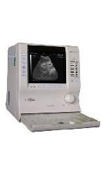 From ALOKA Co., Ltd.;
'Innovative Image Quality in a Compact Size Featuring design concepts inherited from our higher end systems along with Aloka's latest proprietary electronic technologies, the compact SSD-900V delivers superb images. Traditionally, portable units suffer from hardware and software limitations that affect image quality. Aloka's SSD-900V sets new standards for what's possible in a portable unit. Incorporating over 50 years of experience and unique micro-chip technologies, the SSD-900V generates incredibly high-resolution images. The SSD-900V offers a full range of measurement functions for professional ultrasonic examination. And unlike conventional portable systems, the SSD-900V includes annotation labeling and 15-channel preset functions as standard features. The system also use Super High Density transducers (found our high-end systems) to enhance imaging resolution. These multi-frequency transducers provide a broad range of imaging frequencies to optimize scan frequency for depth of view.' • 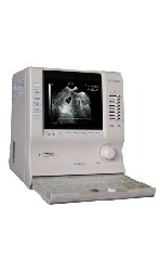 From ALOKA Co., Ltd.;
'Innovative Image Quality in a Compact Size. Featuring design concepts inherited from our higher end systems along with Aloka's latest proprietary electronic technologies, the compact SSD-900 delivers superb images. Traditionally, portable units suffer from hardware and software limitations that affect image quality. Aloka's SSD-900 sets new standards for what's possible in a portable unit. Incorporating over 50 years of experience and unique micro-chip technologies, the SSD-900 generates incredibly high-resolution images. The SSD-900 offers a full range of measurement functions for professional ultrasonic examination. And unlike conventional portable systems, the SSD-900 includes annotation labeling and 15-channel preset functions as standard features. The system also use Super High Density transducers (found our high-end systems) to enhance imaging resolution. These multi-frequency transducers provide a broad range of imaging frequencies to optimize scan frequency for depth of view.' Result Pages : | Share This Page Look Ups |
Medical-Ultrasound-Imaging.com
former US-TIP.com
Member of SoftWays' Medical Imaging Group - MR-TIP • Radiology TIP • Medical-Ultrasound-Imaging
Copyright © 2008 - 2024 SoftWays. All rights reserved.
Terms of Use | Privacy Policy | Advertise With Us
former US-TIP.com
Member of SoftWays' Medical Imaging Group - MR-TIP • Radiology TIP • Medical-Ultrasound-Imaging
Copyright © 2008 - 2024 SoftWays. All rights reserved.
Terms of Use | Privacy Policy | Advertise With Us
[last update: 2023-11-06 01:42:00]