Medical Ultrasound Imaging
Sunday, 12 May 2024
Info Sheets Out- side     | 'Real' p4 Searchterm 'Real' found in 60 articles 3 terms [ • ] - 57 definitions [• ] Result Pages : •
Ultrasound machines, widely used in medical imaging, are essential tools in the field of diagnostic ultrasound. These devices utilize high-frequency sound waves to create real-time images of internal body structures. Ultrasound machines consist of several key components that work together to generate diagnostic images.
These include:
•
The transducer is a handheld device that emits and receives sound waves. It converts electrical energy into sound waves and captures the returning echoes to create images.
•
The control panel houses the interface where the sonographer adjusts imaging parameters such as depth, frequency, and gain. It allows for customization of imaging settings based on the clinical requirements. The transducer pulse controls change the amplitude, frequency and duration of the pulses emitted from the transducer probe.
•
The central processing unit (CPU) serves as the brain of the ultrasound machine, processing the acquired data and transforming it into images. It handles complex calculations, image optimization, data storage and contains the electrical power supplies for itself and the transducer probe.
•
The display monitor (oscilloscope, tablet, computer monitor, etc.) showcases the real-time ultrasound images produced by the machine. It provides visual feedback to the sonographer, aiding in the interpretation and analysis of anatomical structures. Handheld ultrasound devices and mobile ultrasound probes can be connected wirelessly to a smartphone or tablet via Bluetooth or WiFi. These end device serves then as the ultrasound monitor.
•
Data input and measurements are done with the keyboard cursor (trackball). Ultrasound devices used for handheld point of care ultrasound (HPOCUS) are operated via the touch screen of the control panel.
•
Images are captured, reviewed, stored and transmitted digitally, using a standard format for digital imaging and communications in medicine (DICOM). Disk storage devices (FDD, HDD, CD, DVD) are outdated, but may be used in older machines to store the acquired images if no picture archiving and communication system (PACS) connection is possible.
•
The displayed ultrasound pictures are usually digitally stored in a PACS. The images from portable ultrasound machines can be stored and conveniently managed on the end device itself, the inserted memory card or in the cloud. With a QR scanner, the images can be accessed via the Internet in the cloud. Often there is also the possibility to get a picture of a baby sonography as a printout.
B-mode machines represent the vast majority of machines used in echocardiology, obstetrical scans, abdominal scans, gynecological scans, etc. B-mode ultrasound machines usually produce the sector (or pie segment-shaped) scans. These ultrasound scans require either a mechanical scanner transducer (the transducer moves to produce the sector scan), or a linear array transducer operated as a phased array. Ultrasound machines come in different types, each catering to specific clinical needs. The two primary types are stationary and portable ultrasound machines: •
Stationary units are typically larger in size and are installed in dedicated imaging rooms. These machines offer advanced imaging capabilities and a wide range of specialized features. They are commonly found in hospitals, clinics, and university medical centers where comprehensive imaging services are provided.
•
Portable units (see Portable Ultrasound Machine), as the name suggests, are compact and lightweight, designed for on-the-go imaging. These machines are highly versatile and offer excellent mobility, allowing healthcare professionals to bring the ultrasound system directly to the patient's bedside. Portable ultrasound machines are particularly useful in emergency settings, rural healthcare facilities, and point-of-care applications.
See also Handheld Ultrasound, Ultrasound System Performance, Equipment Preparation, Coaxial Cable, and Microbubble Scanner Modification, Environmental Protection and Ultrasound Accessories and Supplies. Further Reading: Basics: News & More:
•
The 2D-mode (2-Dimensional-mode) is a spatially oriented B-mode (brightness) ultrasound. The imaged structures are displayed 2 dimensional as a function of depth and width. The brightness level is based on the echo signal amplitude. Most of the ultrasound devices in medical imaging are 2D real-time scanner. The image is created by a rapidly back and forth swept sound beam over the region of interest. See also Gray Scale. Further Reading: News & More:
•
As far as ultrasound is concerned, 4D ultrasound (also referred to as live 3D ultrasound or 4B-mode) is the latest ultrasound technology - the fourth dimension means length, width, and depth over time. 4D Ultrasound takes 3D ultrasound images and adds the element of time to the progress so that a moving three-dimensional image is seen on the monitor. A 4D scan takes the same amounts of time as a 2D or 3D scan; the difference is the ultrasound equipment being used. One advantage of a 4D fetal ultrasound to a 2D-mode is that parents can see how their baby will generally look like. However, there are different opinions over the medical advantages. To scan a 3D ultrasound image, the probe is swept over the maternal abdomen. A computer takes multiple images and renders the 3D picture. With 4D imaging, the computer takes the images as multiple pictures while the probe is hold still and a 3D image is simultaneously rendered in real time on a monitor. In most cases, the standard 2D ultrasound is taken, and then the 3D/4D scan capability is added if an abnormality is detected or suspected. The 3D/4D sonogram is then focused on a specific area, to provide the details needed to assess and diagnose a suspected problem. A quick 4D scan of the face of the fetus may be performed at the end of a routine exam, providing the parents with a photo. • 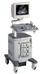 From ALOKA Co., Ltd.;
'A Platform for Digital, Pure-Beam Imaging The high-performance, ALOKA ProSound SSD-3500 utilizes advanced ProSound technologies including: Fully digital beam former A wide dynamic range, 12-bit A/D converter Multi beam processing. The SSD-3500 also helps you achieve more efficient examinations. Its ergonomic, user-friendly design enables you to customize the system according to your specific application needs.'
Device Information and Specification
APPLICATIONS
CONFIGURATION
Compact, portable, dual dynamic display
Color Flow, Power Flow, Spectral Doppler, Real-time Free Angular M-Mode, Tissue Harmonic Imaging, Quint Frequency Imaging, Pure Harmonic Detection
STORAGE, CONNECTIVITY, OS
Data Management Subsystem (iDMS), DICOM-Worklist
DATA PROCESSING
12-bit analog to digital converter
• 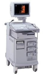 From ALOKA Co., Ltd.;
'The ProSound SSD-4000 utilizes the most advanced acoustic technologies available today, and its multidisciplinary technology architecture enables it to offer great versatility and flexibility over a wide range of clinical applications. With its new-generation, front-end technology including a 12-bit A/D converter, the ProSound SSD-4000 offers superior contrast resolution−especially when compared to 10-bit systems.'
Device Information and Specification
APPLICATIONS
CONFIGURATION
Compact, portable, dual dynamic display
Wide-band super high-density (W-SHD) transducers
OPTIONAL PACKAGE
Volume Mode
STORAGE, CONNECTIVITY, OS
Data Management Subsystem (iDMS), DICOM-Worklist
DATA PROCESSING
Result Pages : | Share This Page Look Ups |
Medical-Ultrasound-Imaging.com
former US-TIP.com
Member of SoftWays' Medical Imaging Group - MR-TIP • Radiology TIP • Medical-Ultrasound-Imaging
Copyright © 2008 - 2024 SoftWays. All rights reserved.
Terms of Use | Privacy Policy | Advertise With Us
former US-TIP.com
Member of SoftWays' Medical Imaging Group - MR-TIP • Radiology TIP • Medical-Ultrasound-Imaging
Copyright © 2008 - 2024 SoftWays. All rights reserved.
Terms of Use | Privacy Policy | Advertise With Us
[last update: 2023-11-06 01:42:00]