Medical Ultrasound Imaging
Monday, 13 May 2024
Info Sheets Out- side     | 'Second' p2 Searchterm 'Second' found in 50 articles 1 term [ • ] - 49 definitions [• ] Result Pages : •
Superharmonic imaging uses higher harmonics like third and fourth harmonic to increase the contrast to tissue ratio compared to a second harmonic imaging mode. Second harmonic imaging is better than fundamental imaging, but has limited capabilities to discriminate between tissue and microbubbles, caused by the non-linear propagation of ultrasound.
•
The traveling ultrasonic wave causes a low-level ultrasound radiation force when this energy is absorbed in tissues (absorbed dose). This force produces a pressure in the direction of the beam and away from the transducer. It should not be confused with the oscillatory pressure of the ultrasound wave itself. The pressure that results and the pressure gradient across the beam are very low, even for intensities at the higher end of the range of diagnostic ultrasound. Mechanical effects like radiation forces lead to stress at tissue interfaces. The effect of the force is manifest in volumes of fluid where streaming can occur with motion within the fluid. The fluid velocities which result are low and are unlikely to cause damage. The effects of ultrasound radiation force (also called Bjerknes Forces) were first reported in 1906 by C. A. and V. F. K. Bjerknes, when they observed the attraction and repulsion of air bubbles in a sound field. While incompressible objects do experience radiation forces, compressible objects driven at their resonant frequency experience far larger forces and can be observably displaced by low-amplitude ultrasound waves. A microbubble driven near its resonance frequency experiences a large net radiation force in the direction of ultrasound wave propagation. Ultrasound pulses of many cycles can deflect resonant microbubbles over distances on the order of millimeters. In addition to primary radiation force, which acts in the direction of acoustic wave propagation, a secondary radiation force for which each individual bubble is a source and receptor causes the microspheres to attract or repel each other. The result of this secondary force is that a much larger concentration of microbubbles collects along a vessel wall than might otherwise occur. See also Acoustically Active Lipospheres. Further Reading: News & More:
•
A-mode (Amplitude-mode) ultrasound is a technique used to assess organ dimensions and determine the depth of an organ. While A-mode technology was previously employed in midline echoencephalography for rapid screening of intracranial mass lesions and ophthalmologic scanning, it is now considered obsolete in medical imaging. Nonetheless, the A-mode scan has found applications in early pregnancy assessment (specifically the detection of fetal heartbeats), cephalometry, and placental localization.
When the ultrasound beam encounters an anatomic boundary, the received sound impulse is processed to appear as a vertical reflection of a point. On the display, it looks like spikes of different heights (the amplitude). The intensity of the returning impulse determined the height of the vertical reflection and the time it took for the impulse to make the round trip would determine the space between verticals. The distance between these spikes can be measured accurately by dividing the speed of sound in tissue (1540 m/sec) by half the sound travel time. During an echoencephalography scan, the first A-mode scan is acquired from the right side of the head and captured on film. Subsequently, the probe is positioned at the corresponding point on the left side, and a second exposure is captured on the same film, displaying inverted spikes. The A-mode ultrasound could be used to identify structures normally located in the midline of the brain such as the third ventricle and falx cerebri. The midline structures would be aligned in normal patients but show displacement in patients with mass lesion such as a subdural, epidural, or intracranial hemorrhage. See also 2D Ultrasound, 3D Ultrasound, 4D Ultrasound, Ultrasound Biomicroscopy, A-scan, B-mode and the Infosheet about ultrasound modes. • 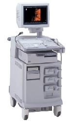 From ALOKA Co., Ltd.;
'The ProSound SSD-4000 utilizes the most advanced acoustic technologies available today, and its multidisciplinary technology architecture enables it to offer great versatility and flexibility over a wide range of clinical applications. With its new-generation, front-end technology including a 12-bit A/D converter, the ProSound SSD-4000 offers superior contrast resolution−especially when compared to 10-bit systems.'
Device Information and Specification
APPLICATIONS
CONFIGURATION
Compact, portable, dual dynamic display
Wide-band super high-density (W-SHD) transducers
OPTIONAL PACKAGE
Volume Mode
STORAGE, CONNECTIVITY, OS
Data Management Subsystem (iDMS), DICOM-Worklist
DATA PROCESSING
•  From ALOKA Co., Ltd.;
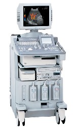
'A Platform for Pure Harmonic Detection The SSD-5000 is the second platform in our ProSound™ line of superior ultrasound systems. The Pure Harmonic Detection technology provides distortion-free fundamental frequency transmission, offering optimum Harmonic Echo™ image quality. This technology is especially effective for obese patients and in a variety of technically difficult scanning conditions.' Result Pages : | Share This Page Look Ups |
Medical-Ultrasound-Imaging.com
former US-TIP.com
Member of SoftWays' Medical Imaging Group - MR-TIP • Radiology TIP • Medical-Ultrasound-Imaging
Copyright © 2008 - 2024 SoftWays. All rights reserved.
Terms of Use | Privacy Policy | Advertise With Us
former US-TIP.com
Member of SoftWays' Medical Imaging Group - MR-TIP • Radiology TIP • Medical-Ultrasound-Imaging
Copyright © 2008 - 2024 SoftWays. All rights reserved.
Terms of Use | Privacy Policy | Advertise With Us
[last update: 2023-11-06 01:42:00]