Medical Ultrasound Imaging
Sunday, 19 May 2024
Info Sheets Out- side     | 'Thyroid Ultrasound' p2 Searchterm 'Thyroid Ultrasound' found in 13 articles 1 term [ • ] - 1 definition [• ] - 11 booleans [• ]Result Pages : •
Shear Waves are waves that travel perpendicular to the direction of the sound beam. During an ultrasound examination, shear waves are generated and transmitted into the body using the ultrasound probe. These waves travel through the tissue and their speed of propagation is influenced by the tissue's stiffness. Softer tissues allow the shear waves to travel faster, while stiffer tissues slow them down.
By analyzing the speed of shear waves, ultrasound systems can provide quantitative measurements of tissue stiffness, known as shear wave elastography. This technique is particularly useful in assessing the stiffness of organs like the liver, breast, thyroid, and muscles.
Shear wave ultrasound elastography has various applications in clinical practice. For example, it can help identify liver fibrosis, a condition characterized by excessive scarring of the liver tissue. By measuring the liver's stiffness using shear waves, clinicians can assess the severity of fibrosis without the need for invasive procedures like liver biopsies.
• View NEWS results for 'Shear Wave' (5). Further Reading: News & More:
•
Sonography [aka: ultrasonography] is a term that encompasses the entire process of performing ultrasound examinations and interpreting the obtained images. Sonography involves the skilled application of ultrasound technology by trained professionals known as sonographers or ultrasound technologists. These specialists operate the ultrasound equipment, manipulate the transducer, and acquire the necessary pictures for diagnostic imaging purposes. Sonography requires in-depth knowledge of anatomy, physiology, and pathology to accurately interpret the ultrasound images and provide valuable information to the treating physician. Sonography uses equipment that generates high frequency sound waves to produce images from muscles, soft tissues, fluid collections, and vascular structures of the human body. Obstetric sonography is commonly used during pregnancy. Sonography visualizes anatomy, function, and pathology of for example gallbladder, kidneys, pancreas, spleen, liver, uterus, ovaries, urinary bladder, eye, thyroid, breast, aorta, veins and arteries in the extremities, carotid arteries in the neck, as well as the heart. A typical medical ultrasound machine, usually a real-time scanner, operates in the frequency range of 2 to 13 megahertz. See also Musculoskeletal and Joint Ultrasound, Pediatric Ultrasound, Cerebrovascular Ultrasonography and Contrast Enhanced Ultrasound. Further Reading: Basics:
News & More:
•
Ultrasound technology with its advancements is vital for delivering high-quality patient care. Innovations including high-frequency ultrasound, 3D//4D imaging, contrast enhanced ultrasound, elastography, and point-of-care ultrasound, have expanded the capabilities of ultrasound imaging and improved diagnostic accuracy. B-Mode imaging, also known as brightness mode, is the fundamental technique in ultrasound imaging. It produces two-dimensional images based on the echoes received from tissues and organs. Understanding the principles of B-Mode imaging, such as gain adjustment, depth control, and image optimization, is crucial for obtaining diagnostically valuable images. M-Mode imaging, on the other hand, allows for the visualization of motion over time, enabling assessment of cardiac structures and function, as well as fetal heart rate. High-frequency ultrasound refers to the use of ultrasound waves with frequencies greater than 10 MHz. This technology enables improved resolution, allowing for detailed imaging of superficial structures like skin, tendons, and small organs. High-frequency ultrasound has found applications in dermatology, ophthalmology, and musculoskeletal imaging. Traditional 2D ultrasound has been augmented by the advent of 3D ultrasound technology. By acquiring multiple 2D images from different angles, this technique construct a volumetric representation of the imaged area. The addition of 4D ultrasound in real-time motion adds further value by capturing dynamic processes. Doppler imaging employs the Doppler effect to evaluate blood flow within vessels and assess hemodynamics. Color Doppler assigns color to different blood flow velocities, providing a visual representation of blood flow direction and speed. Spectral Doppler displays blood flow velocities as a waveform, allowing for detailed analysis of flow patterns, resistance, and stenosis. Contrast enhanced ultrasound employs microbubble contrast agents to enhance the visualization of blood flow and tissue perfusion. By injecting these agents intravenously, sonographers can differentiate between vascular structures and lesions. Elastography is a technique that measures tissue elasticity or stiffness. It assists in differentiating between normal and abnormal tissues, aiding in the diagnosis of various conditions such as liver fibrosis, breast lesions, and thyroid nodules. Fusion imaging combines ultrasound with other imaging modalities, such as computed tomography (CT), magnetic resonance imaging (MRI), or positron emission tomography (PET). By overlaying or merging ultrasound images with those obtained from other modalities, the user can precisely locate and characterize abnormalities, guide interventions, and improve diagnostic accuracy. Fusion imaging has proven particularly useful in areas such as interventional radiology, oncology, and urology. See also Equipment Preparation, Environmental Protection, Handheld Ultrasound, Portable Ultrasound and Ultrasound Accessories and Supplies. •
(US) Also called echography, sonography, ultrasonography, echotomography, ultrasonic tomography. Diagnostic imaging plays a vital role in modern healthcare, allowing medical professionals to visualize internal structures of the body and assist in the diagnosis and treatment of various conditions. Two terms that are commonly used interchangeably but possess distinct meanings in the field of medical imaging are 'ultrasound' and 'sonography.' Ultrasound is the imaging technique that utilizes sound waves to create real-time images, while sonography encompasses the entire process of performing ultrasound examinations and interpreting the obtained images. Ultrasonography is a synonymous term for sonography, emphasizing the use of ultrasound technology in diagnostic imaging. A sonogram, on the other hand, refers to the resulting image produced during an ultrasound examination. Ultrasonic waves, generated by a quartz crystal, cause mechanical perturbation of an elastic medium, resulting in rarefaction and compression of the medium particles. These waves are reflected at the interfaces between different tissues due to differences in their mechanical properties. The transmission and reflection of these high-frequency waves are displayed with different types of ultrasound modes. By utilizing the speed of wave propagation in tissues, the time of reflection information can be converted into distance of reflection information. The use of higher frequencies in medical ultrasound imaging yields better image resolution. However, higher frequencies also lead to increased absorption of the sound beam by the medium, limiting its penetration depth. For instance, higher frequencies (e.g., 7.5 MHz) are employed to provide detailed imaging of superficial organs like the thyroid gland and breast, while lower frequencies (e.g., 3.5 MHz) are used for abdominal examinations. Ultrasound in medical imaging offers several advantages including:
•
noninvasiveness;
•
safety with no potential risks;
•
widespread availability and relatively low cost.
Diagnostic ultrasound imaging is generally considered safe, with no adverse effects. As medical ultrasound is extensively used in pregnancy and pediatric imaging, it is crucial for practitioners to ensure its safe usage. Ultrasound can cause mechanical and thermal effects in tissue, which are amplified with increased output power. Consequently, guidelines for the safe use of ultrasound have been issued to address the growing use of color flow imaging, pulsed spectral Doppler, and higher demands on B-mode imaging. Furthermore, recent ultrasound safety regulations have shifted more responsibility to the operator to ensure the safe use of ultrasound. See also Skinline, Pregnancy Ultrasound, Obstetric and Gynecologic Ultrasound, Musculoskeletal and Joint Ultrasound, Ultrasound Elastography and Prostate Ultrasound. Further Reading: Basics: News & More:
•  From Siemens Medical Systems;
'Conventional ultrasound systems are unable to compensate for the unique acoustic signature of individual patients. But there is nothing conventional about the new ACUSON Sequoia™ C512 echocardiography system with Sequoia™ matched response technology.'
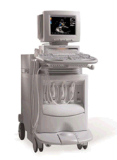
Device Information and Specification
APPLICATIONS
CONFIGURATION
Compact, portable
2D-Mode, M-mode, Cadence™, Native® Tissue Harmonic Imaging, Transmit Compounding, SST™ Color and Solo™ Spectral Doppler
STORAGE, CONNECTIVITY, OS
KinetDx integrated PACS, DIMAQ-Workstation
Result Pages : | Share This Page Look Ups |
Medical-Ultrasound-Imaging.com
former US-TIP.com
Member of SoftWays' Medical Imaging Group - MR-TIP • Radiology TIP • Medical-Ultrasound-Imaging
Copyright © 2008 - 2024 SoftWays. All rights reserved.
Terms of Use | Privacy Policy | Advertise With Us
former US-TIP.com
Member of SoftWays' Medical Imaging Group - MR-TIP • Radiology TIP • Medical-Ultrasound-Imaging
Copyright © 2008 - 2024 SoftWays. All rights reserved.
Terms of Use | Privacy Policy | Advertise With Us
[last update: 2023-11-06 01:42:00]