Medical Ultrasound Imaging
Sunday, 19 May 2024
Info Sheets Out- side     | 'Ultrasound Technology' p2 Searchterm 'Ultrasound Technology' found in 36 articles 1 term [ • ] - 18 definitions [• ] - 17 booleans [• ]Result Pages : •
As far as ultrasound is concerned, 4D ultrasound (also referred to as live 3D ultrasound or 4B-mode) is the latest ultrasound technology - the fourth dimension means length, width, and depth over time. 4D Ultrasound takes 3D ultrasound images and adds the element of time to the progress so that a moving three-dimensional image is seen on the monitor. A 4D scan takes the same amounts of time as a 2D or 3D scan; the difference is the ultrasound equipment being used. One advantage of a 4D fetal ultrasound to a 2D-mode is that parents can see how their baby will generally look like. However, there are different opinions over the medical advantages. To scan a 3D ultrasound image, the probe is swept over the maternal abdomen. A computer takes multiple images and renders the 3D picture. With 4D imaging, the computer takes the images as multiple pictures while the probe is hold still and a 3D image is simultaneously rendered in real time on a monitor. In most cases, the standard 2D ultrasound is taken, and then the 3D/4D scan capability is added if an abnormality is detected or suspected. The 3D/4D sonogram is then focused on a specific area, to provide the details needed to assess and diagnose a suspected problem. A quick 4D scan of the face of the fetus may be performed at the end of a routine exam, providing the parents with a photo. •
Handheld ultrasound systems are portable devices for smartphone or tablet and are increasingly common in emergency, intensive care and veterinary medicine, but also in the pocket of the stationary doctor. This type of ultrasound machine enables immediate diagnoses directly on site (handheld point-of-care-ultrasound / HPOCUS) and quickly provide information regarding the patient's further care. Handheld ultrasound machines fit into a single-use plastic cover and can be easily disinfected, making them particularly useful in infectious environments. The most striking advantage of handheld POCUS devices is the small footprint. The design is very compact, lightweight (approx. 200 g/0,44 lbs. - 500 g/1,1 lbs) and flexible. Due to this compactness and the necessary technical compression, the quality of the imaging is still limited compared to 'high-end devices', but sufficient to the extent that handheld ultrasound devices are already successfully used in many medical disciplines. Depending on the model, handheld ultrasound systems run on Android, iOS, Windows or proprietary operating systems. They are connected to the end device via USB cable or wirelessly via Bluetooth or WiFi. The respective end device is used as an ultrasound monitor to display the ultrasound images. The associated app is operated via touchscreen, although some devices have a few buttons, e.g. for recording ultrasound images or freeze images. The images can be stored and managed on the end device itself, the inserted memory card or in the cloud. Theoretically, also a private smartphone can be connected, but this can lead to complications with reimbursement. See also Portable Ultrasound Machine, Ultrasound Technology, Environmental Protection, Ultrasound Accessories and Supplies and Sonographer. Further Reading: News & More:
•
The earliest introduction of vascular ultrasound contrast agents (USCA) was by Gramiak and Shah in 1968, when they injected agitated saline into the ascending aorta and cardiac chambers during echocardiographic to opacify the left heart chamber. Strong echoes were produced within the heart, due to the acoustic mismatch between free air microbubbles in the saline and the surrounding blood. The disadvantage of this microbubbles produced by agitation, was that the air quickly leak from the thin bubble shell into the blood, where it dissolved. In addition, the small bubbles that were capable of traversing the capillary bed did not survive long enough for imaging because the air quickly dissipated into the blood. Aside from agitated saline, also hydrogen peroxide, indocyanine green dye, and iodinated contrast has been tested. The commercial development of contrast agents began in the 1980s with greatest effort to the stabilization of small microbubbles. The development generations by now:
•
first generation USCA = non-transpulmonary vascular;;
•
second generation USCA = transpulmonary vascular, with short half-life (less than 5 min);
•
third generation USCA = transpulmonary vascular, with longer half-life (greater than 5 min).
To pass through the lung capillaries and enter into the systemic circulation, microspheres should be less than 10 μm in diameter. Air bubbles in that size range persist in solution for only a short time; too short for systemic vascular use. The first developed agent was Echovist (1982), which enabled the enhancement of the right heart. The second generation of echogenic agents, sonicated 5% human albumin-containing air bubbles (Albunex), were capable of transpulmonary passage but often failed to produce adequate imaging of the left heart. Both Albunex and Levovist utilize air as the gas component of the microbubble. In the 1990s newer developed agents with fluorocarbon gases and albumin, surfactant, lipid, or polymer shells have an increased persistence of the microspheres. This smaller, more stable microbubble agents, and improvements in ultrasound technology, have resulted in a wider range of application including myocardial perfusion. See also First Generation USCA, Second Generation USCA, and Third Generation USCA. Further Reading: Basics:
• 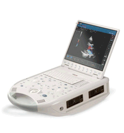 From ESAOTE S.p.A.;
'The MyLab™30CV ultrasound system is an evolutionary step in ultrasound technology. Weighing less than 20 pounds, it is the first compact ultrasound system to deliver premium console performance. And with mobile, portable or stationary configurations, MyLab30CV can adapt to any clinical environment.'
Device Information and Specification
APPLICATIONS
Abdominal, breast, cardiac, OB/GYN, pediatric, pediatric cardiology, small parts, transcranial, vascular
CONFIGURATION
Portable
Linear: 4-10 MHz, convex: 2-5 MHz, phased: 1.6-10 MHz, micro convex: 5-7.5 MHz, endocavity: 5-7.5 MHz, pencil: 2 + 5 MHz
2-D, M-mode, duplex, triplex, color Doppler, pulsed wave Doppler, tissue velocity mapping (TVM), tissue enhancement imaging (TEI™), contrast harmonic imaging, stress echo, tissue velocity mapping for LV motion analysis (TVM), contrast tuned imaging for contrast media procedures (CnTI™), Qontrast™ for myocardium parameters quantification
STORAGE, CONNECTIVITY, OS
Digital patient archive/management, integrated CD/RW, RJ 45 and USB ports, Windows
H*W*D m (inch.)
0.16 * 0.36 * 0.50 (6.2 x 14 x 19.3)
WEIGHT
Less than 11 kg (20 lbs.)
•
As far as ultrasound is concerned, 4D ultrasound (also referred to as live 3D ultrasound or 4B-mode) is the latest ultrasound technology - the fourth dimension means length, width, and depth over time. 4D Ultrasound takes 3D ultrasound images and adds the element of time to the progress so that a moving three-dimensional image is seen on the monitor. A 4D scan takes the same amounts of time as a 2D or 3D scan; the difference is the ultrasound equipment being used. One advantage of a 4D fetal ultrasound to a 2D-mode is that parents can see how their baby will generally look like. However, there are different opinions over the medical advantages. To scan a 3D ultrasound image, the probe is swept over the maternal abdomen. A computer takes multiple images and renders the 3D picture. With 4D imaging, the computer takes the images as multiple pictures while the probe is hold still and a 3D image is simultaneously rendered in real time on a monitor. In most cases, the standard 2D ultrasound is taken, and then the 3D/4D scan capability is added if an abnormality is detected or suspected. The 3D/4D sonogram is then focused on a specific area, to provide the details needed to assess and diagnose a suspected problem. A quick 4D scan of the face of the fetus may be performed at the end of a routine exam, providing the parents with a photo. See also Obstetric and Gynecologic Ultrasound, Pregnancy Ultrasound, Fetal Ultrasound and Abdominal Ultrasound. Result Pages : | Share This Page Look Ups |
Medical-Ultrasound-Imaging.com
former US-TIP.com
Member of SoftWays' Medical Imaging Group - MR-TIP • Radiology TIP • Medical-Ultrasound-Imaging
Copyright © 2008 - 2024 SoftWays. All rights reserved.
Terms of Use | Privacy Policy | Advertise With Us
former US-TIP.com
Member of SoftWays' Medical Imaging Group - MR-TIP • Radiology TIP • Medical-Ultrasound-Imaging
Copyright © 2008 - 2024 SoftWays. All rights reserved.
Terms of Use | Privacy Policy | Advertise With Us
[last update: 2023-11-06 01:42:00]