Medical Ultrasound Imaging
Tuesday, 14 May 2024
Info Sheets Out- side     | 'probe' p20 Searchterm 'probe' found in 121 articles 11 terms [ • ] - 110 definitions [• ] Result Pages : •
Ultrasound is an ideal tool to examine the joints and surrounding soft tissues like tendons, ligaments and joint linings. Musculoskeletal and joint sonography is sensitive, without radiation exposure, easy accessible, quick, and has high patient tolerability with relatively low cost. A real-time scanner allow the dynamic assessment of the musculoskeletal system and a specific examination for each patient. In addition, joint aspiration and injection accuracy can be improved. Probes with high frequency improve the image resolution and allow visualization of fine anatomic structures of the small parts. As musculoskeletal ultrasound (MSUS) is very operator dependent, experience and training is required. Ultrasound is also often used in the treatment of musculoskeletal disorders. See also Ultrasound Therapy, Real-Time Mode, Artifact and Ultrasound Biomicroscopy. Further Reading: Basics:
• 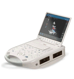 From ESAOTE S.p.A.;
'The MyLab™30CV ultrasound system is an evolutionary step in ultrasound technology. Weighing less than 20 pounds, it is the first compact ultrasound system to deliver premium console performance. And with mobile, portable or stationary configurations, MyLab30CV can adapt to any clinical environment.'
Device Information and Specification
APPLICATIONS
Abdominal, breast, cardiac, OB/GYN, pediatric, pediatric cardiology, small parts, transcranial, vascular
CONFIGURATION
Portable
Linear: 4-10 MHz, convex: 2-5 MHz, phased: 1.6-10 MHz, micro convex: 5-7.5 MHz, endocavity: 5-7.5 MHz, pencil: 2 + 5 MHz
2-D, M-mode, duplex, triplex, color Doppler, pulsed wave Doppler, tissue velocity mapping (TVM), tissue enhancement imaging (TEI™), contrast harmonic imaging, stress echo, tissue velocity mapping for LV motion analysis (TVM), contrast tuned imaging for contrast media procedures (CnTI™), Qontrast™ for myocardium parameters quantification
STORAGE, CONNECTIVITY, OS
Digital patient archive/management, integrated CD/RW, RJ 45 and USB ports, Windows
H*W*D m (inch.)
0.16 * 0.36 * 0.50 (6.2 x 14 x 19.3)
WEIGHT
Less than 11 kg (20 lbs.)
•
The propagation of high amplitude ultrasound waves is inadequate described by a linear wave equation. Non-linear propagation is to expect if the power levels are high enough to make non-linear effects significant.
A non-linear propagation results in the distortion of the transmitted waveforms, resulting in the generation of harmonics of the initial frequency components transmitted by the transducer. In the near field of ultrasound probes, the occurring diffraction and focusing effects make this process complex. The distortion of a wavefront propagating in a medium in which the compressional phase moves slightly faster than the rarefactional phase, results is the conversion of some wave energy into higher harmonics of the fundamental frequency. The effect increases strongly with increasing wave amplitude. Further Reading: News & More:
• •
Pregnancy ultrasound plays a crucial role in monitoring the health and development of the fetus throughout pregnancy. It serves as a screening tool with various applications, including:
•
Verification of Due Date and Assessment of Pregnancy Health: Fetal ultrasound examinations are used to accurately determine the estimated due date of the baby. They also aid in investigating the causes of bleeding during pregnancy and assessing the overall health and well-being of the fetus. •
•
Measurement of Amniotic Fluid and Placental Assessment: Ultrasound is utilized to measure the amniotic fluid levels, which provide insights into fetal well-being and the functioning of the placenta. It also helps evaluate the condition of the placenta, ensuring proper nutrient and oxygen supply to the developing baby. •
Early Pregnancy Confirmation and Multiple Fetuses Detection: Around week five to seven of pregnancy, ultrasound is utilized to confirm the pregnancy, determine the fetal size, and detect the presence of multiple fetuses. It aids in distinguishing between intrauterine and ectopic pregnancies, ensuring appropriate management. •
Third-Trimester Evaluation: As the pregnancy progresses, ultrasound assessments are conducted to evaluate fetal size, position, growth, and the condition of the placenta. This information assists healthcare providers in monitoring the well-being of the fetus and planning for a safe delivery. •
Guiding Procedures: Ultrasound plays a vital role in guiding invasive procedures such as amniocentesis and chorionic villus sampling. It helps guide the placement of a needle to collect cells from the amniotic fluid or placenta, aiding in genetic testing and diagnosing potential fetal abnormalities. See also Doppler Fluximetry in Pregnancy, Fetal Ultrasound, Obstetric and Gynecologic Ultrasound and Vaginal Probe. Result Pages : | Share This Page Look Ups |
Medical-Ultrasound-Imaging.com
former US-TIP.com
Member of SoftWays' Medical Imaging Group - MR-TIP • Radiology TIP • Medical-Ultrasound-Imaging
Copyright © 2008 - 2024 SoftWays. All rights reserved.
Terms of Use | Privacy Policy | Advertise With Us
former US-TIP.com
Member of SoftWays' Medical Imaging Group - MR-TIP • Radiology TIP • Medical-Ultrasound-Imaging
Copyright © 2008 - 2024 SoftWays. All rights reserved.
Terms of Use | Privacy Policy | Advertise With Us
[last update: 2023-11-06 01:42:00]