Medical Ultrasound Imaging
Sunday, 19 May 2024
Info Sheets Out- side     | '2D Ultrasound' p4 Searchterm '2D Ultrasound' found in 20 articles 1 term [ • ] - 5 definitions [• ] - 14 booleans [• ]Result Pages : •
Unlike regular sound, ultrasound can be directed into a single direction. The echoes received by a stationary probe will result in a single dimensional signal showing peaks for every major material change. To generate a 2D picture, the probe is swiveled, either mechanically or through a phased array of ultrasound transducers. The data is analyzed by computer and used to construct the image. In a similar way, 3D pictures can be generated by computer using a specialized probe. In this way, a photo of an unborn baby may be made. Some ultrasonography machines can produce color pictures, of sorts. Doppler ultrasonography is color coded onto a gray scale picture. From the amount of energy in each echo, the difference in acoustic impedance can be calculated and a color is then assigned accordingly. See also Densitometry and 3D Ultrasound. •
(CD) Color Doppler is an ultrasound imaging mode, which visualizes the presence, direction and velocity of flowing blood in a wide range of flow conditions. It provides an estimate of the mean velocity of flow within a vessel by color coding the flow and displaying it superimposed on the 2D gray scale image. The flow direction is arbitrarily assigned the color red or blue, indicating flow toward or away from the transducer. Color (colour, Brit.) Doppler ultrasound is capable of evaluating a wider area than other Doppler modes than for example Duplex or power Doppler, and therefore makes it less likely to miss flow abnormalities. It is also easier to interpret. Color flow is not as precise as conventional Doppler and is best used to scan a larger area and then use conventional Doppler for detailed analysis at a site of potential flow abnormality. Adjustments for color Doppler in case of too much color: Adjustments for color Doppler in case of not enough color:
•
increased color gain;
•
decrease color velocity scale;
•
adjust scanning plane and angle to flow;
•
decrease sample box size;
•
evaluation of chosen filter.
See also Color Power Doppler, Autocorrelation, Color Priority, Triplex Exam and Color Saturation. Further Reading: Basics:
News & More:
•
Also called B-mode echography, B-mode sonography, 2D-mode, and sonogram. B-mode ultrasound (Brightness-mode) is the display of a 2D-map of B-mode data, currently the most common form of ultrasound imaging. The development from A-mode to B-mode is that the ultrasound signal is used to produce various points whose brightness depends on the amplitude instead of the spiking vertical movements in the A-mode. Sweeping a narrow ultrasound beam through the area being examined while transmitting pulses and detecting echoes along closely spaced scan lines produces B-scan images. The vertical position of each bright dot is determined by the time delay from pulse transmission to return of the echo, and the horizontal position by the location of the receiving transducer element. To generate a rapid series of individual 2D images that show motion, the ultrasound beam is swept repeatedly. The returning sound pulses in B-mode have different shades of darkness depending on their intensities. The varying shades of gray reflect variations in the texture of internal organs. This form of display (solid areas appear white and fluid areas appear black) is also called gray scale. Different types of displayed B-mode images are: The probe movement can be performed manual (compound and static B-scanner) or automatic (real-time scanner). The image reconstruction can be parallel or sector type. See also B-Scan, 4B-Mode, and Harmonic B-Mode Imaging. Further Reading: News & More:
•
Doppler ultrasound is a medical imaging technique for calculating the relative velocity between two points by measuring the frequency shift of a sound wave transmitted from one point to the other, based on the Doppler effect. Continuous or pulsed Doppler is frequently used to examine cardiovascular blood flow. The combination of routine 2D-mode and Doppler ultrasound allows a complete evaluation of the heart's anatomy and function (including the fetal heart). See also Doppler Fluximetry in Pregnancy. Doppler ultrasound depends on the fact that if a moving object reflects the ultrasound waves, the echo frequencies are changed. A higher frequency is created if the object is moving toward the probe//transducer and a lower frequency if it is moving away from it. How much the frequency is changed depends upon how fast the object is moving. Doppler ultrasound shows the different rates of blood flow in different colors on a monitor in real time. The major Doppler parameters are the peak systolic velocity and the end-diastolic velocity. The peak systolic velocity ratio compensates the variability between different patients and instrumentations. Different Doppler and duplex techniques: Further Reading: News & More:
• 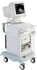 From Biosound Esaote, Inc.;
'The Caris Plus combines the power of a conventional mainframe ultrasound system with an extraordinarily portable design. The result − High-performance ultrasound diagnostics in the most compact, most portable CFM system available anywhere. Powerful state-of-the-art microelectronics and wide-band multi-frequency technology ensure exceptional 2D, Doppler and CFM performance that can be taken where they're needed.' Specifications for this system will be available soon. Result Pages : | Share This Page Look Ups |
Medical-Ultrasound-Imaging.com
former US-TIP.com
Member of SoftWays' Medical Imaging Group - MR-TIP • Radiology TIP • Medical-Ultrasound-Imaging
Copyright © 2008 - 2024 SoftWays. All rights reserved.
Terms of Use | Privacy Policy | Advertise With Us
former US-TIP.com
Member of SoftWays' Medical Imaging Group - MR-TIP • Radiology TIP • Medical-Ultrasound-Imaging
Copyright © 2008 - 2024 SoftWays. All rights reserved.
Terms of Use | Privacy Policy | Advertise With Us
[last update: 2023-11-06 01:42:00]