Medical Ultrasound Imaging
Sunday, 19 May 2024
Info Sheets Out- side     | 'Pediatric Ultrasound' p4 Searchterm 'Pediatric Ultrasound' found in 20 articles 1 term [ • ] - 6 definitions [• ] - 13 booleans [• ]Result Pages : •  From SIUI Inc.;
'Dedicated to ultrasound industry, Shantou Institute of Ultrasonic Instruments, Inc. (SIUI) has launched Apogee 3500, the Digital Color Doppler Ultrasound Imaging System. With latest imaging technologies, high-definition image quality and excellent practical functions, the Apogee 3500 offers optimal solutions for clinical ultrasonic examination.' 'The Apogee 3500 is available with many high-density, super broadband and multi-frequency probes, such as convex, micro-convex, linear, vaginal, rectal and phased array probes, which are widely applied for different clinical diagnoses, including abdomen (liver, kidney, gall-bladder, pancreas), gynecology (uterus, ovary), obstetrics (early pregnancy, basic OB, complete OB, multi gestation, fetal echo), cardiology (adult and pediatric cardiology), small parts (thyroid, galactophore, testicles, neonate), peripheral vascular and prostate.'
Device Information and Specification
CONFIGURATION
Normal system, color - gray scale(256)
Linear, convex and phased array
PROBES STANDARD
2.0 MHz ~ 12.0 MHz, broad band, tri-frequency
B-mode (B, 2B, 4B), M-mode, B/M-mode, real-time compound imaging, panoramic imaging, trapezoidal imaging (linear probes), spectrum Doppler (PWD and CWD), color Doppler flow imaging (CDFI), color power angio (CPA), tissue harmonic imaging (THI)
IMAGING OPTIONS
Real-time ZOOM, zoom rate and position selectable
OPTIONAL PACKAGE
H*W*D m
1.29 * 0.52 * 0.75
WEIGHT
110 kg
POWER REQUIREMENT
AC 220V/110V, 50Hz/60Hz
POWER CONSUMPTION
0.6 KVA
•  From SIUI Inc.;
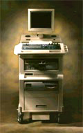
'The Apogee 800Plus ultrasound system offers a new level of high quality in image. With broad clinical application, the system delivers ease of use for cardiac and vascular exams.' 'Due to the exceptional image quality, the sensitivity of color Doppler and many other advanced features, the Apogee 800Plus offers complete diagnosis capability for your entire patient population, including abdominal, obstetrics, gynecology, adult cardiology, pediatric cardiology, peripheral vascular, small parts, breast, prostate, transcranial, etc.'
Device Information and Specification
APPLICATIONS
Linear, curved, phased and symmetric phased array
2D, M-mode, Pulsed wave Doppler, Color Power Angio Imaging, Color 2D, Color M-mode, High PRF Doppler
H*W*D m
1.46 * 0.69 * 0.87
WEIGHT
140 kg (without peripherals)
POWER REQUIREMENT
115Vac, (V~), @ 50 to 60 Hz, 1.2kVA
230Vac, (V~), @ 50 to 60 Hz, 1.2kVA POWER CONSUMPTION
800 Watts (with optional OEM'S: 1200 Watts max)
•
Lateral resolution is the minimum separation of two interfaces aligned along a direction perpendicular (objects that are side by side) to the ultrasound beam. The lateral or angular resolution directly relates with the collimation of the beam emitted by the crystal. Lateral resolution is proportionally affected by the frequency, the higher the frequency the greater the lateral resolution.
Higher frequency transducers are used in fetal and pediatric echocardiography because the lateral resolution displays the smaller structures better. Lower frequencies are used for adults where structures are larger and the need for greater depth penetration is important. Further Reading: Basics: •
(US) Also called echography, sonography, ultrasonography, echotomography, ultrasonic tomography. Diagnostic imaging plays a vital role in modern healthcare, allowing medical professionals to visualize internal structures of the body and assist in the diagnosis and treatment of various conditions. Two terms that are commonly used interchangeably but possess distinct meanings in the field of medical imaging are 'ultrasound' and 'sonography.' Ultrasound is the imaging technique that utilizes sound waves to create real-time images, while sonography encompasses the entire process of performing ultrasound examinations and interpreting the obtained images. Ultrasonography is a synonymous term for sonography, emphasizing the use of ultrasound technology in diagnostic imaging. A sonogram, on the other hand, refers to the resulting image produced during an ultrasound examination. Ultrasonic waves, generated by a quartz crystal, cause mechanical perturbation of an elastic medium, resulting in rarefaction and compression of the medium particles. These waves are reflected at the interfaces between different tissues due to differences in their mechanical properties. The transmission and reflection of these high-frequency waves are displayed with different types of ultrasound modes. By utilizing the speed of wave propagation in tissues, the time of reflection information can be converted into distance of reflection information. The use of higher frequencies in medical ultrasound imaging yields better image resolution. However, higher frequencies also lead to increased absorption of the sound beam by the medium, limiting its penetration depth. For instance, higher frequencies (e.g., 7.5 MHz) are employed to provide detailed imaging of superficial organs like the thyroid gland and breast, while lower frequencies (e.g., 3.5 MHz) are used for abdominal examinations. Ultrasound in medical imaging offers several advantages including:
•
noninvasiveness;
•
safety with no potential risks;
•
widespread availability and relatively low cost.
Diagnostic ultrasound imaging is generally considered safe, with no adverse effects. As medical ultrasound is extensively used in pregnancy and pediatric imaging, it is crucial for practitioners to ensure its safe usage. Ultrasound can cause mechanical and thermal effects in tissue, which are amplified with increased output power. Consequently, guidelines for the safe use of ultrasound have been issued to address the growing use of color flow imaging, pulsed spectral Doppler, and higher demands on B-mode imaging. Furthermore, recent ultrasound safety regulations have shifted more responsibility to the operator to ensure the safe use of ultrasound. See also Skinline, Pregnancy Ultrasound, Obstetric and Gynecologic Ultrasound, Musculoskeletal and Joint Ultrasound, Ultrasound Elastography and Prostate Ultrasound. Further Reading: Basics: News & More:
• Verathon Inc. formerly 'Diagnostic Ultrasound' was founded in 1984 by Gerald McMorrow. His vision was to use emerging ultrasound technology to develop noninvasive, easy to use medical devices that would fill unmet needs in health care. In the beginning, the company was no more than one man working alone in his basement. Today, the company has 150 employees and has sold more than 100 million dollars worth of its products worldwide. Ultrasound Systems:
Contact Information
ONLINE
CONTACT
Result Pages : | Share This Page Look Ups |
Medical-Ultrasound-Imaging.com
former US-TIP.com
Member of SoftWays' Medical Imaging Group - MR-TIP • Radiology TIP • Medical-Ultrasound-Imaging
Copyright © 2008 - 2024 SoftWays. All rights reserved.
Terms of Use | Privacy Policy | Advertise With Us
former US-TIP.com
Member of SoftWays' Medical Imaging Group - MR-TIP • Radiology TIP • Medical-Ultrasound-Imaging
Copyright © 2008 - 2024 SoftWays. All rights reserved.
Terms of Use | Privacy Policy | Advertise With Us
[last update: 2023-11-06 01:42:00]