Medical Ultrasound Imaging
Sunday, 20 September 2026

 Info Sheets  Out- side     | 'SD' Searchterm 'SD' found in 199 articles 26 terms [ • ] - 173 definitions [• ] Result Pages : •
(Perfluorocarbon exposed sonicated dextrose albumin) PESDA are sonicated dextrose albumin microbubbles containing decafluorobutane gas. PESDA is a sonicated self made contrast agent and not a commercial product. PESDA microbubbles can be used as a blood pool contrast agent in echocardiography to improve the detection of myocardial perfusion abnormalities.
Drug Information and Specification
RESEARCH NAME
-
DEVELOPER
-
INDICATION -
DEVELOPMENT STAGE APPLICATION
Intravenous injection
TYPE
Microbubble
Albumin / dextrose
CHARGE
-
Decafluorobutane
MICROBUBBLE SIZE
Mean: 4.7 ± 0.2 μm
PREPARATION
-
DO NOT RELY ON THE INFORMATION PROVIDED HERE, THEY ARE
NOT A SUBSTITUTE FOR THE ACCOMPANYING PACKAGE INSERT!  Further Reading: Basics:
News & More: • 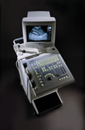 From Philips Medical Systems;
'The powerful system... The SD 260 is a powerful B/W ultrasound system that offers standard all major scan techniques: curved, linear and annular phased array. The user has a choice of no less than 12 probes and can connect up to three probes simultaneously, regardless of their technique. The SD 260 is perfectly suited for use in either a busy hospital or clinic.' This product is not available in the US, Australia, or New Zealand from Philips Medical Systems. Specifications for this system will be available soon. • 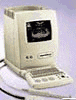 From Philips Medical Systems;
'The perfect practice system... The SD 240E is a compact and ergonomically designed system, standard equipped with two probe connectors. The user has a choice out of more than 12 linear, curved and annular phased array probes which can be used with this perfect office unit. Total Image Focus and cine memory technology are just the icing on the cake.' This product is not available in the US, Australia, or New Zealand from Philips Medical Systems.' Specifications for this system will be available soon. •
A transducer is a device, usually electrical or electronic, that converts one type of energy to another. Most transducers are either sensors or actuators. A transducer (also called probe) is a main part of the ultrasound machine. The transducer sends ultrasound waves into the body and receives the echoes produced by the waves when it is placed on or over the body part being imaged. Ultrasound transducers are made from crystals with piezoelectric properties. This material vibrates at a resonant frequency, when an alternating electric current is applied. The vibration is transmitted into the tissue in short bursts. The speed of transmission within most soft tissues is 1540 m/s, producing a transit time of 6.5 ms/cm. Because the velocity of ultrasound waves is constant, the time taken for the wave to return to the transducer can be used to determine the depth of the object causing the reflection. The waves will be reflected when they encounter a boundary between two tissues of different density (e.g. soft tissue and bone) and return to the transducer. Conversely, the crystals emit electrical currents when sound or pressure waves hit them (piezoelectric effect). The same crystals can be used to send and receive sound waves; the probe then acts as a receiver, converting mechanical energy back into an electric signal which is used to display an image. A sound absorbing substance eliminates back reflections from the probe itself, and an acoustic lens focuses the emitted sound waves. Then, the received signal gets processed by software to an image which is displayed at a monitor. Transducer heads may contain one or more crystal elements. In multi-element probes, each crystal has its own circuit. The advantage is that the ultrasound beam can be controlled by changing the timing in which each element gets pulsed. Especially for cardiac ultrasound it is important to steer the beam. Usually, several different transducer types are available to select the appropriate one for optimal imaging. Probes are formed in many shapes and sizes. The shape of the probe determines its field of view. Transducers are described in megahertz (MHz) indicating their sound wave frequency. The frequency of emitted sound waves determines how deep the sound beam penetrates and the resolution of the image. Most transducers are only able to emit one frequency because the piezoelectric ceramic or crystals within it have a certain inherent frequency, but multi-frequency probes are also available. See also Blanking Distance, Damping, Maximum Response Axis, Omnidirectional, and Huygens Principle. Further Reading: News & More:
• 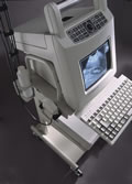 From Philips Medical Systems;
'A perfect portable system... The SD 100 is a system designed to travel. Weighing only 10.5 kg and battery or mains operated, the SD 100E is a full-featured ultrasound unit with a comprehensive software package, nine-inch monitor, digital output and built-in diskette for software updates and image storage. The SD 100E user can choose from seven linear and convex array dual frequency probes to perform many ultrasound examinations, quickly and reliably.' This product is not available in the US, Australia, or New Zealand from Philips Medical Systems. Specifications for this system will be available soon. Result Pages : | Share This Page     Look Ups |
 (.pdf)
Medical-Ultrasound-Imaging.com
former US-TIP.com
Member of SoftWays' Medical Imaging Group - MR-TIP • Radiology TIP • Medical-Ultrasound-Imaging
Copyright © 2008 - 2025 SoftWays. All rights reserved.
Terms of Use | Privacy Policy | Advertise With Us
former US-TIP.com
Member of SoftWays' Medical Imaging Group - MR-TIP • Radiology TIP • Medical-Ultrasound-Imaging
Copyright © 2008 - 2025 SoftWays. All rights reserved.
Terms of Use | Privacy Policy | Advertise With Us
[last update: 2025-04-05 02:38:00]