Medical Ultrasound Imaging
Wednesday, 8 May 2024
Info Sheets Out- side     | 'Transducer Types' Searchterm 'Transducer Types' found in 17 articles 1 term [ • ] - 2 definitions [• ] - 14 booleans [• ]Result Pages : • Transducer Types
Transducers can be divided in: 1.) Transducers where the sound wave is transmitted and received by different elements. 2.) Transducers where multiple elements part of the time transmit and part of the time receive sound energy. The first type of ultrasound transducer is used in detection of blood flow (also called nonimaging transducers). For example, the continuous wave transducer (Pedoff transducer) has two separate elements, where one element is always transmitting while the other element is always receiving. Probes of the second type are used to image cardiac structures and have the capability to use various Doppler techniques to detect blood flow (also called imaging transducers). For example, continuous wave, pulsed wave, high pulse repetition frequency, color flow, M-mode, and 2D-mode are the various modes that this type of transducer can perform. Transducers can also be divided in mechanical and electronic or phased scan types. Mechanical transducers use a combination of single element oscillation, multiple element rotation, or a single element and set of acoustic mirrors to generate the sweeping beam for 2D mode. Caused by the vibration (created as the mirrors rotate or oscillate inside the cover) is this type sometimes called the 'wobbler'. Mechanical transducers are cheaper than electronic transducers. Different types of electronic or phased array probes can create a linear or rectangular shaped scan plane as well as a sector or pie shaped scan plane. Sector scanners are most useful for cardiac ultrasound examinations where the beam is directed between the ribs to image the heart. A linear array transducer is more useful in abdominal, OB/GYN, and small parts examinations. Electronic transducers are more expensive but they provide dynamic focusing and smaller probe. See also Rectangular Array Transducer. •
An array transducer is composed of multiple piezoelectric crystal elements arranged in an array. Arrays are transducer assemblies with a row of elements, used to focus the beam. Types of array transducers:
•
Linear array transducer = the arrays are arranged along a line.
•
Curvilinear or curved transducer = the arrays are arranged along a convex curve. A curved array is similar to a linear array except that the image created is a sector-type.
•
Annular array transducer = the arrays are arranged in concentric circles.
•
Rectangular array transducer = the arrays are arranged in a rectangular pattern.
See also Amplitude Shading, Transducer Types, and Transducer Assembly. Further Reading: Basics:
•
A transducer is a device, usually electrical or electronic, that converts one type of energy to another. Most transducers are either sensors or actuators. A transducer (also called probe) is a main part of the ultrasound machine. The transducer sends ultrasound waves into the body and receives the echoes produced by the waves when it is placed on or over the body part being imaged. Ultrasound transducers are made from crystals with piezoelectric properties. This material vibrates at a resonant frequency, when an alternating electric current is applied. The vibration is transmitted into the tissue in short bursts. The speed of transmission within most soft tissues is 1540 m/s, producing a transit time of 6.5 ms/cm. Because the velocity of ultrasound waves is constant, the time taken for the wave to return to the transducer can be used to determine the depth of the object causing the reflection. The waves will be reflected when they encounter a boundary between two tissues of different density (e.g. soft tissue and bone) and return to the transducer. Conversely, the crystals emit electrical currents when sound or pressure waves hit them (piezoelectric effect). The same crystals can be used to send and receive sound waves; the probe then acts as a receiver, converting mechanical energy back into an electric signal which is used to display an image. A sound absorbing substance eliminates back reflections from the probe itself, and an acoustic lens focuses the emitted sound waves. Then, the received signal gets processed by software to an image which is displayed at a monitor. Transducer heads may contain one or more crystal elements. In multi-element probes, each crystal has its own circuit. The advantage is that the ultrasound beam can be controlled by changing the timing in which each element gets pulsed. Especially for cardiac ultrasound it is important to steer the beam. Usually, several different transducer types are available to select the appropriate one for optimal imaging. Probes are formed in many shapes and sizes. The shape of the probe determines its field of view. Transducers are described in megahertz (MHz) indicating their sound wave frequency. The frequency of emitted sound waves determines how deep the sound beam penetrates and the resolution of the image. Most transducers are only able to emit one frequency because the piezoelectric ceramic or crystals within it have a certain inherent frequency, but multi-frequency probes are also available. See also Blanking Distance, Damping, Maximum Response Axis, Omnidirectional, and Huygens Principle. Further Reading: News & More:
• 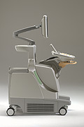 From Philips Medical Systems;
'The Philips iU22 system combines Intelligent Design, including breakthroughs in ergonomics, with Intelligent Control, providing new levels of automation, to give you revolutionary performance and workflow.'
Device Information and Specification
APPLICATIONS
Abdominal, cardiac (also for adults with TEE), musculoskeletal (also pediatric), OB/GYN, prostate, smallparts, transcranial, vascular
CONFIGURATION
17' high resolution non-interlaced flat CRT, 4 active probe ports
B-mode, M-mode, coded harmonic imaging, color flow mode (CFM), power Doppler imaging (PDI), color Doppler, pulsed wave Doppler, tissue harmonic imaging
IMAGING OPTIONS
CrossXBeam spatial compounding, coded ultrasound acquisition),speckle reduction imaging (SRI), TruScan technology store raw data, CINE review with 4 speed types
OPTIONAL PACKAGE
Transesophageal scanning, stress echo, tissue velocity imaging (TVI), tissue velocity Doppler (TVD), contrast harmonic imaging
STORAGE, CONNECTIVITY, OS
Patient and image archive, HDD, DICOM 3.0, CD/DVD, MOD, Windows-based
DATA PROCESSING
Digital beamformer with 1024 system processing channel technology
H*W*D m (inch.)
1.62 * 0.61 * 0.99 (64 * 22 * 43)
WEIGHT
kg (345 lbs.)
POWER CONSUMPTION
•
Cardiac ultrasound, also known as echocardiography or echocardiogram, is used to provide several different levels and types of heart testing. Cardiac ultrasound utilizes the same ultrasound principles as used for obstetric and gynecologic evaluations of pregnant women, gallbladder ultrasound and other abdominal structures. The ultrasound is directed out of a hand held probe which can be moved to image the heart from different positions. Additionally, so that heart events can be timed, ECG leads are placed on the chest. The reflected wave is converted into an actual image of the heart and displayed in a real-time mode or M-mode ultrasound format. M-mode recordings permit measurement of cardiac dimensions and detailed analysis of complex motion patterns depending on transducer angulations. Also the time relationships with other physiological variables such as ECG, heart sounds, and pulse tracings, can be recorded simultaneously. A stress echocardiogram provides information about the cardiac performance. Two-dimensional tomographic images of selected cardiac sections give more information than M-mode about the shape of the heart and also show the spatial relationships of its structures during the cardiac cycle (diastole to systole). See also M-Mode Echocardiography, and Myocardial Contrast Echocardiography. Further Reading: News & More:
Result Pages : | Share This Page Look Ups |
Medical-Ultrasound-Imaging.com
former US-TIP.com
Member of SoftWays' Medical Imaging Group - MR-TIP • Radiology TIP • Medical-Ultrasound-Imaging
Copyright © 2008 - 2024 SoftWays. All rights reserved.
Terms of Use | Privacy Policy | Advertise With Us
former US-TIP.com
Member of SoftWays' Medical Imaging Group - MR-TIP • Radiology TIP • Medical-Ultrasound-Imaging
Copyright © 2008 - 2024 SoftWays. All rights reserved.
Terms of Use | Privacy Policy | Advertise With Us
[last update: 2023-11-06 01:42:00]