Medical Ultrasound Imaging
Wednesday, 8 May 2024
Info Sheets Out- side     | 'Dynamic Focusing' Searchterm 'Dynamic Focusing' found in 7 articles 1 term [ • ] - 3 definitions [• ] - 3 booleans [• ]Result Pages : • Dynamic Focusing
Dynamic focusing controls the axial position of the focus of an ultrasound beam. Dynamic focusing is often managed by controlling the phase of the signals detected by a transducer array. See also Real-Time Transducer. • 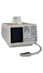 From ALOKA Co., Ltd.;
'Veterinary portable ultrasound system: The SSC-210 Vet is a price performance breakthrough in diagnostic imaging. The variable frame rate, up to 30 frames/sec., combined with dynamic focusing produces highly detailed images that can be optimized and frozen at the touch of a button. The push key controlled, linear distance calipers permit fast, accurate measurement of any structure within the two-dimensional image. The built-in digital scan converter let you output the image directly to a video recording system.' •
Transducers used for the real-time mode are different than for the A-mode, B-, or M-modes. A linear array transducer with multiple piezoelectric crystal elements that are different arranged and fired, transmits the needed larger sound beam. A subgroup of x adjacent elements (8-16; or more in wide-aperture designs) is pulsed simultaneously; the inner elements pulse delayed with respect to the outer elements. The interference of the x small divergent wavelets generates a focused beam. The delay time determining the focus depth of a real-time transducer can be changed during imaging. Similar delay factors applied during the receiving phase, result in a dynamic focusing effect on the return. This forms a single scan line in the real-time image. To produce the following scan line, another group of x elements is selected by shifting one element position along the transducer array from the previous group. This pattern is then repeated for the groups along the array, in a sequential and repetitive way. Further Reading: Basics: •
Transducers can be divided in: 1.) Transducers where the sound wave is transmitted and received by different elements. 2.) Transducers where multiple elements part of the time transmit and part of the time receive sound energy. The first type of ultrasound transducer is used in detection of blood flow (also called nonimaging transducers). For example, the continuous wave transducer (Pedoff transducer) has two separate elements, where one element is always transmitting while the other element is always receiving. Probes of the second type are used to image cardiac structures and have the capability to use various Doppler techniques to detect blood flow (also called imaging transducers). For example, continuous wave, pulsed wave, high pulse repetition frequency, color flow, M-mode, and 2D-mode are the various modes that this type of transducer can perform. Transducers can also be divided in mechanical and electronic or phased scan types. Mechanical transducers use a combination of single element oscillation, multiple element rotation, or a single element and set of acoustic mirrors to generate the sweeping beam for 2D mode. Caused by the vibration (created as the mirrors rotate or oscillate inside the cover) is this type sometimes called the 'wobbler'. Mechanical transducers are cheaper than electronic transducers. Different types of electronic or phased array probes can create a linear or rectangular shaped scan plane as well as a sector or pie shaped scan plane. Sector scanners are most useful for cardiac ultrasound examinations where the beam is directed between the ribs to image the heart. A linear array transducer is more useful in abdominal, OB/GYN, and small parts examinations. Electronic transducers are more expensive but they provide dynamic focusing and smaller probe. See also Rectangular Array Transducer. •
Linear array transducer elements are rectangular and arranged in a line. Linear array probes are described by the radius of width in mm. A linear array transducer can have up to 512 elements spaced over 75-120 mm. The beam produced by such a narrow element will diverge rapidly after the wave travels only a few millimeters. The smaller the face of the transducer, the more divergent is the beam. This would result in poor lateral resolution due to beam divergence and low sensitivity due to the small element size. In order to overcome this, adjacent elements are pulsed simultaneously (typically 8 to 16; or more in wide-aperture designs). In a subgroup of x elements, the inner elements pulse delayed with respect to the outer elements. The interference of the x small divergent wavelets produces a focused beam. The delay time determines the depth of focus for the transmitted beam and can be changed during scanning. Linear arrays are usually cheaper than sector scanners but have greater skin contact and therefore make it difficult to reach organs between ribs such as the heart. One-dimensional linear array transducers may have dynamic, electronic focusing providing a narrow ultrasound beam in the image plane. In the z-plane (elevation plane - perpendicular to the image plane) focusing may be provided by an acoustic lens with a fixed focal zone. Rectangular or matrix transducers with unequal rows of transducer elements are two-dimensional (2D), but they are termed 1.5D, because the number of rows is much less than the number of columns. These transducers provide dynamic, electronic focusing even in the z-plane. See also Rectangular Array Transducer. Result Pages : | Share This Page Look Ups |
Medical-Ultrasound-Imaging.com
former US-TIP.com
Member of SoftWays' Medical Imaging Group - MR-TIP • Radiology TIP • Medical-Ultrasound-Imaging
Copyright © 2008 - 2024 SoftWays. All rights reserved.
Terms of Use | Privacy Policy | Advertise With Us
former US-TIP.com
Member of SoftWays' Medical Imaging Group - MR-TIP • Radiology TIP • Medical-Ultrasound-Imaging
Copyright © 2008 - 2024 SoftWays. All rights reserved.
Terms of Use | Privacy Policy | Advertise With Us
[last update: 2023-11-06 01:42:00]