Medical Ultrasound Imaging
Sunday, 19 May 2024
Info Sheets Out- side     | 'Contrast Harmonic Imaging' p3 Searchterm 'Contrast Harmonic Imaging' found in 24 articles 1 term [ • ] - 8 definitions [• ] - 15 booleans [• ]Result Pages : • 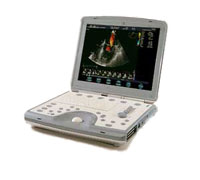 From GE Healthcare.;
'The incredible Vivid i system establishes a completely new level of cardiovascular performance that gives clinicians the freedom to get diagnostic results outside of the echo lab.'
Device Information and Specification
APPLICATIONS
CONFIGURATION
Notebook
M-mode (and 2-D), triplex mode, harmonic imaging, color flow mapping, pulsed wave Doppler, continuous wave Doppler, power Doppler, color Doppler, tissue harmonic imaging, color flow mapping
IMAGING OPTIONS
STORAGE, CONNECTIVITY, OS
Patient and image archive, HDD, DICOM, CD/DVD, MOD, USB flash, PCMCIA, eVue for remote monitoring, MPEGvue foruniversal record sharing
H*W*D cm (inch.)
7 * 36 * 32 (2.6 x 14.1 x 12.3)
WEIGHT
5 kg (11 lbs.)
POWER CONSUMPTION
Rechargeable battery provides up to 1.0 hour of full scan operation
• View NEWS results for 'Vivid i' (1). •
(CPS) Contrast pulse sequencing is a technique to exploit contrast agent properties with series of three pulses that differ in phase and amplitude. CPS allows bubble specific imaging with non-linear fundamental and higher order harmonics, low MI, and extremely high microbubble-to-tissue background ratio. See also Ultrasound Contrast Agent Safety. •
(LVO) Ultrasound contrast agents improve the echocardiography assessment of left ventricular function and the low sensitivity of changes in left ventricular ejection fraction (LVEF). In addition, harmonic imaging techniques and automated border detection (ABD) together with contrast enhanced left ventricular opacification increase endocardial border delineation (EBD) and the results compared to native echocardiography.
Further Reading: Basics:
•
(PII) Pulse inversion imaging (also called phase inversion imaging) is a non-linear imaging method specifically made for enhanced detection of microbubble ultrasound contrast agents. In PII, two pulses are sent in rapid succession into the tissue; the second pulse is a mirror image of the first. The resulting echoes are added at reception. Linear scattering of the two pulses will give two echoes which are inverted copies of each other, and these echoes will therefore cancel out when added. Linear scattering dominates in tissues. Echoes from linear scatterers such as tissue cancel, whereas those from gas microbubbles do not. Non-linear scattering of the two pulses will give two echoes which do not cancel out completely due to different bubble response to positive and negative pressures of equal magnitude. The harmonic components add, and the signal intensity difference between non-linear and linear scatterers is therefore increased. The resulting images show high sensitivity to bubbles at the resolution of a conventional image. In harmonic imaging, the frequency range of the transmitted pulse and the received signal should not overlap, but this restriction is less in pulse inversion imaging since the transmit frequencies are not filtered out, but rather subtracted. Broader transmit and receive bandwidths are therefore allowed, giving shorter pulses and improved axial resolution, hence the alternative term wideband harmonic imaging. Many ultrasound machines offer some form of pulse inversion imaging. See also Pulse Inversion Doppler, Narrow Bandwidth, Dead Zone, Ultrasound Phantom. •
Conventional, CT and MR imaging technologies are limited in their availability, to depict soft tissue, or to show dynamic activity, like cardiac muscle contractility and blood flow. Easy applicability, real-time sonography and biopsy facilitation are important advantages in veterinarian medicine. Veterinary ultrasound has a very high sensitivity to show the composition of soft tissues, but the low specificity is a disadvantage. High ultrasound system performance includes Doppler techniques, contrast enhanced ultrasound, 3D ultrasound, and tissue harmonic imaging to improve resolution. Technical and physical requirements of veterinary ultrasound are the same as in human ultrasonography. The higher the sound frequency, the better the possible resolution, but the poorer the tissue penetration. Image quality is depended of the ultrasound equipment. For example, a 10 MHz transducer is excellent for imaging of superficial structures; a 3.5 or 5.0 megahertz transducer allows sufficient penetration to see inner structures like the liver or the heart. In addition, the preparation and performing of the examination is similar to that of humans. The sound beam penetrates soft tissue and fat well, but gas and bone impede the ultrasonic power. Fluid filled organs like the bladder are often used as an acoustic window, and an ultrasound gel is used to conduct the sound beam. Result Pages : | Share This Page Look Ups |
Medical-Ultrasound-Imaging.com
former US-TIP.com
Member of SoftWays' Medical Imaging Group - MR-TIP • Radiology TIP • Medical-Ultrasound-Imaging
Copyright © 2008 - 2024 SoftWays. All rights reserved.
Terms of Use | Privacy Policy | Advertise With Us
former US-TIP.com
Member of SoftWays' Medical Imaging Group - MR-TIP • Radiology TIP • Medical-Ultrasound-Imaging
Copyright © 2008 - 2024 SoftWays. All rights reserved.
Terms of Use | Privacy Policy | Advertise With Us
[last update: 2023-11-06 01:42:00]