Medical Ultrasound Imaging
Sunday, 19 May 2024
Info Sheets Out- side     | 'Curved Transducer' p2 Searchterm 'Curved Transducer' found in 12 articles 1 term [ • ] - 4 definitions [• ] - 7 booleans [• ]Result Pages : • 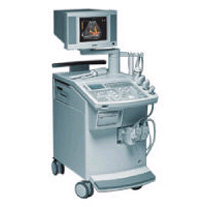 From Siemens Medical Systems;
'This extremely flexible system supports targeted applications for OB/GYN, Radiology, Internal Medicine, Urology, and Prostate Brachytherapy. The large transducer selection, most with high-frequency capabilities, provides the right tool for general imaging, radiology, and internal medicine applications.'
Device Information and Specification
CLINICAL APPLICATION
OB/GYN, cerebrovascular, orthopedics, urology, peripheral vascular, pediatrics, small parts, surgery, abdominal, musculoskeletal
CONFIGURATION
Mobile, compact
DISPLAY MODES
Broadband, high-fidelity, multi-frequency, linear and curved
PROBE PORTS
Two - four
256
MEASUREMENT/CALCULATION FUNCTIONS
OB/GYN measurements and report package, off-line analysis
OPTIONAL PACKAGE
IMAGE PROCESSING
• 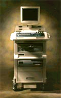 From SIUI Inc.;
'The Apogee 800Plus ultrasound system offers a new level of high quality in image. With broad clinical application, the system delivers ease of use for cardiac and vascular exams.' 'Due to the exceptional image quality, the sensitivity of color Doppler and many other advanced features, the Apogee 800Plus offers complete diagnosis capability for your entire patient population, including abdominal, obstetrics, gynecology, adult cardiology, pediatric cardiology, peripheral vascular, small parts, breast, prostate, transcranial, etc.'
Device Information and Specification
APPLICATIONS
Linear, curved, phased and symmetric phased array
2D, M-mode, Pulsed wave Doppler, Color Power Angio Imaging, Color 2D, Color M-mode, High PRF Doppler
H*W*D m
1.46 * 0.69 * 0.87
WEIGHT
140 kg (without peripherals)
POWER REQUIREMENT
115Vac, (V~), @ 50 to 60 Hz, 1.2kVA
230Vac, (V~), @ 50 to 60 Hz, 1.2kVA POWER CONSUMPTION
800 Watts (with optional OEM'S: 1200 Watts max)
•
Most usual ultrasound machines are 2D real-time systems. This types of ultrasound scanners allow to assess both motion and anatomy, including the motion of heart valves, the movement of intestines and lungs and also to guide interventions, like for example a biopsy or a laparoscopic ultrasound. A standard real-time scanner consists of a mobile console with the monitor on the top and rows of small containers at the bottom to accommodate a variety of scanner probes. The linear, curved or phased array transducers are usually equipped with multiple crystals or in some cases with a moving crystal. A real-time scanner may be e.g., a mechanical scanner or electronic array scanner. See also Musculoskeletal and Joint Ultrasound. Further Reading: News & More: •
Sonography of the gallbladder is a reliable technique for diagnosing e.g., gallstones, cholecystitis, tumors, polyps, or ductal obstruction.
Patient should be examined with empty stomach and on a low fat diet the night before. Barium studies, endoscopy, ERCP, colonoscopy, and abdominal CT should be performed after this examination. Gallbladder ultrasound is best performed with a 5 MHz curved array or a linear array transducer in cases of a very superficial gallbladder. In obese patients or in patients with difficult sonographic access, a 3.5 MHz sector or curved linear transducer is advantageous. Gallbladder and biliary tree are usually imaged in supine and posterior oblique (LPO) positions. Sometimes very small gallstones are better visible in upright and prone position. Further Reading: Basics:
News & More:
•
The array of elements of microconvex probe is curved with a certain radius.
Microconvex probes have a much smaller contact surface, which improves the coupling between the transducer and the skin surface even in complicated areas as the supraclavicular or jugular fossa. Microconvex probes, with large aperture and selection of transmission frequencies are also used in gynecological diagnostic. See also Transvaginal Echography, Endocavitary Echography and Transrectal Ultrasonography. Result Pages : | Share This Page Look Ups |
Medical-Ultrasound-Imaging.com
former US-TIP.com
Member of SoftWays' Medical Imaging Group - MR-TIP • Radiology TIP • Medical-Ultrasound-Imaging
Copyright © 2008 - 2024 SoftWays. All rights reserved.
Terms of Use | Privacy Policy | Advertise With Us
former US-TIP.com
Member of SoftWays' Medical Imaging Group - MR-TIP • Radiology TIP • Medical-Ultrasound-Imaging
Copyright © 2008 - 2024 SoftWays. All rights reserved.
Terms of Use | Privacy Policy | Advertise With Us
[last update: 2023-11-06 01:42:00]