Medical Ultrasound Imaging
Tuesday, 14 May 2024
Info Sheets Out- side     | 'Device' p3 Searchterm 'Device' found in 67 articles 1 term [ • ] - 66 definitions [• ] Result Pages : •
Ultrasound machines, with their various components and types, have revolutionized the field of medical imaging. These devices enable healthcare professionals to visualize internal structures, assess conditions, and guide interventions with real-time imaging capabilities.
Today, medical ultrasound systems are complex signal processing machines. Assessing the performance of an ultrasound system requires understanding the relationships between the characteristics of the system, such as the point spread function, temporal resolution, and the quality of images. Image quality aspects include the detail resolution, contrast resolution and penetration. Systems with microbubble scanner modification are particularly suitable for contrast enhanced ultrasound.
•
Low-performance systems constitute approximately 20% of the world ultrasound market. These ultrasound machines are characterized by basic black and white imaging and are primarily used for basic OB/GYN applications and fetal development monitoring. They are often purchased by private office practitioners and small hospitals, with a unit cost below $50,000. These scanners commonly come equipped with a transvaginal probe.
•
Mid-performance sonography systems also hold around 20% market share. These machines are basic gray scale imaging, color and spectral Doppler and are used for routine examinations and reporting. They typically utilize a minimum number of scanheads and find applications in radiology, cardiology, and OB/GYN. The cost of these systems ranges between $50,000 and $100,000. Refurbished advanced and high-performance ultrasound machines with fewer optional features can also be found in this price range.
•
High-performance ultrasound systems generally provide high-resolution gray scale imaging, advanced color power and spectral Doppler capabilities. They usually include advanced measurement and analysis software, image review capabilities, and a variety of probes. These high-performance sonography devices have a market share of approximately 40% and cost between $100,000 and $150,000.
•
The remaining 20% of the market consists of premium or advanced performance ultrasound systems, typically sold for over $150,000. Premium performance systems offer high-resolution gray scale imaging, advanced color flow, power Doppler, and spectral Doppler, as well as features like tissue harmonic imaging, image acquisition storage, display and review capabilities, advanced automation, and more. Premium systems are equipped with a wide assortment of transducer scanheads.
In summary, ultrasound machines have diverse performance levels and corresponding price ranges, catering to various medical imaging needs. From low-performance systems with basic imaging capabilities to high-performance and premium systems with advanced features, ultrasound technology continues to advance healthcare imaging capabilities. See also Ultrasound Physics, Handheld Ultrasound, Environmental Protection, Equipment Preparation. Further Reading: Basics:
•
2D ultrasound imaging is a widely used technique in medical imaging that provides two-dimensional visual representations of internal structures. A handheld device known as a probe or transducer contains piezoelectric crystals that emit and receive ultrasound waves which penetrate tissues and bounce back as echoes. The echoes are detected and converted into electrical signals. These signals are processed and displayed on a monitor, creating a real-time 2D grayscale image, with different shades of gray representing various tissue densities. The brighter areas on the image correspond to structures that reflect more ultrasound waves, while darker areas represent structures that reflect fewer waves or are attenuated by intervening tissues. The 2D-mode (or B-mode) provides cross-sectional views of the scanned area, showing a single plane or slice of the scanned area at a time. Key Features and Uses of 2D Ultrasound: •
•
2D ultrasound is excellent for visualizing anatomical structures and detecting anomalies. It is widely used in obstetrics, gynecology, abdominal imaging and vascular examinations.
•
Due to its real-time capabilities, 2D ultrasound is utilized to guide various procedures, including biopsies, injections, and catheter insertions.
•
2D sonography can incorporate Doppler technology to assess blood flow in vessels, aiding in the diagnosis of vascular conditions and evaluating fetal circulation.
Comparison with 3D and 4D Ultrasound: •
Unlike 2D ultrasound, which generates a series of 2D images, 3D ultrasound creates a three-dimensional volume of the scanned area. This allows for more detailed visualization of complex structures, such as fetal facial features or organ morphology.
•
4D ultrasound adds the dimension of time to 3D imaging, resulting in dynamic three-dimensional videos. It enables the visualization of fetal movements and provides a more immersive experience. However, a 4D sonogram is not typically used for diagnostic purposes and is often employed in baby ultrasound examinations for bonding and enjoyment purposes.
See also Ultrasound Technology, Sonographer, Ultrasound Elastography, Obstetric and Gynecologic Ultrasound. •
The 2D-mode (2-Dimensional-mode) is a spatially oriented B-mode (brightness) ultrasound. The imaged structures are displayed 2 dimensional as a function of depth and width. The brightness level is based on the echo signal amplitude. Most of the ultrasound devices in medical imaging are 2D real-time scanner. The image is created by a rapidly back and forth swept sound beam over the region of interest. See also Gray Scale. Further Reading: News & More:
• 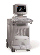 From Siemens Medical Systems;
'Conventional ultrasound systems are unable to compensate for the unique acoustic signature of individual patients. But there is nothing conventional about the new ACUSON Sequoia™ C512 echocardiography system with Sequoia™ matched response technology.'
Device Information and Specification
APPLICATIONS
CONFIGURATION
Compact, portable
2D-Mode, M-mode, Cadence™, Native® Tissue Harmonic Imaging, Transmit Compounding, SST™ Color and Solo™ Spectral Doppler
STORAGE, CONNECTIVITY, OS
KinetDx integrated PACS, DIMAQ-Workstation
• 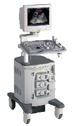 From ALOKA Co., Ltd.;
'A Platform for Digital, Pure-Beam Imaging The high-performance, ALOKA ProSound SSD-3500 utilizes advanced ProSound technologies including: Fully digital beam former A wide dynamic range, 12-bit A/D converter Multi beam processing. The SSD-3500 also helps you achieve more efficient examinations. Its ergonomic, user-friendly design enables you to customize the system according to your specific application needs.'
Device Information and Specification
APPLICATIONS
CONFIGURATION
Compact, portable, dual dynamic display
Color Flow, Power Flow, Spectral Doppler, Real-time Free Angular M-Mode, Tissue Harmonic Imaging, Quint Frequency Imaging, Pure Harmonic Detection
STORAGE, CONNECTIVITY, OS
Data Management Subsystem (iDMS), DICOM-Worklist
DATA PROCESSING
12-bit analog to digital converter
Result Pages : | Share This Page Look Ups |
Medical-Ultrasound-Imaging.com
former US-TIP.com
Member of SoftWays' Medical Imaging Group - MR-TIP • Radiology TIP • Medical-Ultrasound-Imaging
Copyright © 2008 - 2024 SoftWays. All rights reserved.
Terms of Use | Privacy Policy | Advertise With Us
former US-TIP.com
Member of SoftWays' Medical Imaging Group - MR-TIP • Radiology TIP • Medical-Ultrasound-Imaging
Copyright © 2008 - 2024 SoftWays. All rights reserved.
Terms of Use | Privacy Policy | Advertise With Us
[last update: 2023-11-06 01:42:00]