Medical Ultrasound Imaging
Sunday, 19 May 2024
Info Sheets Out- side     | 'Enhancement' p3 Searchterm 'Enhancement' found in 28 articles 2 terms [ • ] - 26 definitions [• ] Result Pages : • 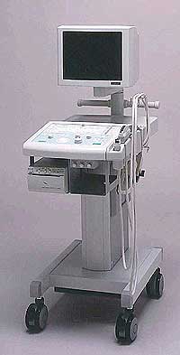 From Fukuda Denshi Co., Ltd.;
'Operation-friendly Switches and Controls backlit to permit the operator to identify them in a darkroom Image Optimization through Ultrasonic Frequency Changeover without replacement of the probe High frame rate enhancement mode for Cardiac echo Cine memory for selection of best data through SCROLL, LOOP, or MANUAL review Programmable function keys allow to get predefined for measurement parameters. Special measurement software usable in a wide range. Measurement Report page on the display can be printed as required.' •
Gray scale [also grayscale, grey scale = brit.] produces basically black and white images with series of shades of gray. Solid areas appear white and fluid areas appear black, varying from black at the weakest intensity to white at the strongest. Gray scale resolves artifacts as small as 1 mm. The display is made by transmitting bursts of energy and analyzing the returning signal. Gray scale pictures are limited to the gray scale tones; color pictures display more information because the color is added to the gray scale. Most ultrasound contrast agents also improve gray scale visualization of the flowing blood to such a degree that the tissue echogenicity increases. Gray scale enhancement of flow in an organ promises to improve lesion detection, along with the ability to differentiate between normal and abnormal areas, using many of the criteria already routinely used in CT and MRI. See also Compress, Densitometry, Triplex Exam and QB-Mode. •  From Hitachi Medical Corporation (HMC), sales, marketing and service in the US by Hitachi Medical Systems America Inc.;
Powerful, flexible, and fast, the HI VISION™ 8500 - EUB-8500 diagnostic ultrasound scanner combines leading edge technologies with user-oriented operation for exceptional imaging and functionality. Available exclusively on the 8500, SonoElastography provides a new perspective on the physical properties of tumors and masses by determining and displaying the relative stiffness of tissue. Device Information and Specification
APPLICATIONS
Abdominal, brachytherapy/cryotherapy, breast, cardiac, dedicated biopsy, endoscopic, intraoperative, laparoscopic, musculoskeletal, OB/GYN, pediatric, small parts, urologic, vascular
CONFIGURATION
Compact system
RANGE OF PROBE TYPE
Linear, convex, radial, biplane, phased array, echoendoscope longitudinal, echoendoscope radial
PROBE FREQUENCIES
Linear: 5.0-13 MHz, convex: 2.5-7.5 MHz, phased:
2.0-7.5 MHz, sector: 2.0-7.5 MHz
4 Modes of dynamic tissue harmonic imaging (dTHI), pulsed wave Doppler, continuous wave Doppler, color flow imaging, power Doppler, directional power Doppler, color flow angiography, real-time Doppler measurements, quantitative tissue Doppler
IMAGING OPTIONS
HI COMPOUND imaging,
HI RES adaptive imaging, wideband pulse inversion imaging (WPI), Raw Data Freeze
OPTIONAL PACKAGE
IMAGING ENHANCEMENTS
3RD generation color artifact suppression
STORAGE, CONNECTIVITY, OS
Patient and image database management system, HDD, FDD, MOD, CD-ROM, Network, DICOM 3.0, Windows XP
DATA PROCESSING
Octal beam processing, 12 bit Gigasampling A/D for precise signal reproduction
H*W*D m (inch.)
1.50 * 0.56 * 1.02 (59 x 22 x 40)
WEIGHT
159 kg (351 lbs.)
POWER CONSUMPTION
1.5kVA
•
The earliest introduction of vascular ultrasound contrast agents (USCA) was by Gramiak and Shah in 1968, when they injected agitated saline into the ascending aorta and cardiac chambers during echocardiographic to opacify the left heart chamber. Strong echoes were produced within the heart, due to the acoustic mismatch between free air microbubbles in the saline and the surrounding blood. The disadvantage of this microbubbles produced by agitation, was that the air quickly leak from the thin bubble shell into the blood, where it dissolved. In addition, the small bubbles that were capable of traversing the capillary bed did not survive long enough for imaging because the air quickly dissipated into the blood. Aside from agitated saline, also hydrogen peroxide, indocyanine green dye, and iodinated contrast has been tested. The commercial development of contrast agents began in the 1980s with greatest effort to the stabilization of small microbubbles. The development generations by now:
•
first generation USCA = non-transpulmonary vascular;;
•
second generation USCA = transpulmonary vascular, with short half-life (less than 5 min);
•
third generation USCA = transpulmonary vascular, with longer half-life (greater than 5 min).
To pass through the lung capillaries and enter into the systemic circulation, microspheres should be less than 10 μm in diameter. Air bubbles in that size range persist in solution for only a short time; too short for systemic vascular use. The first developed agent was Echovist (1982), which enabled the enhancement of the right heart. The second generation of echogenic agents, sonicated 5% human albumin-containing air bubbles (Albunex), were capable of transpulmonary passage but often failed to produce adequate imaging of the left heart. Both Albunex and Levovist utilize air as the gas component of the microbubble. In the 1990s newer developed agents with fluorocarbon gases and albumin, surfactant, lipid, or polymer shells have an increased persistence of the microspheres. This smaller, more stable microbubble agents, and improvements in ultrasound technology, have resulted in a wider range of application including myocardial perfusion. See also First Generation USCA, Second Generation USCA, and Third Generation USCA. Further Reading: Basics:
•
Imagent® is an injectable suspension for intravenous administration during a sonogram. This diagnostic contrast agent for enhancement of ultrasound images contains perflexane lipid microspheres. Imagent® US is indicated in the assessment of heart function and perfusion, as well as the detection of tumors and blood flow abnormalities by using gray scale, color Doppler, and harmonic ultrasound imaging techniques. During the course of its development, the brand name for this product has changed from Imagent to Imavist (between August 2000 and March 2002) back to Imagent. The manufacturer's 06/03/02 press release announcing FDA approval refers to the product as 'Imagent (formerly Imavist),' and the approval notice and monograph posted at the FDA site refers to the product as Imagent. Jointly developed by Alliance Pharmaceutical Corp and Bayer Schering Pharma AG (Germany). Source: PR Newswire - 10/10/96, 03/31/98, 10/13/99, 03/13/01, 10/08/01; FDA approvals - 05/31/02; Alliance Pharmaceutical press release - 06/03/02. Currently the production of Imagent® is discontinued.
Drug Information and Specification
RESEARCH NAME
AF0150
DEVELOPER
IMCOR Pharmaceuticals, Inc.
INDICATION
APPLICATION
Intravenous
TYPE
Lipid: DMPC
CHARGE
Neutral
Perfluorohexane/Nitrogen
MICROBUBBLE SIZE
99.8% < 10μm
PRESENTATION
-
STORAGE
Room Temp 15−30 °C
PREPARATION
Reconstitute with 10 ml water
DO NOT RELY ON THE INFORMATION PROVIDED HERE, THEY ARE
NOT A SUBSTITUTE FOR THE ACCOMPANYING PACKAGE INSERT! Result Pages : | Share This Page Look Ups |
Medical-Ultrasound-Imaging.com
former US-TIP.com
Member of SoftWays' Medical Imaging Group - MR-TIP • Radiology TIP • Medical-Ultrasound-Imaging
Copyright © 2008 - 2024 SoftWays. All rights reserved.
Terms of Use | Privacy Policy | Advertise With Us
former US-TIP.com
Member of SoftWays' Medical Imaging Group - MR-TIP • Radiology TIP • Medical-Ultrasound-Imaging
Copyright © 2008 - 2024 SoftWays. All rights reserved.
Terms of Use | Privacy Policy | Advertise With Us
[last update: 2023-11-06 01:42:00]