Medical Ultrasound Imaging
Monday, 20 May 2024
Info Sheets Out- side     | 'Frame Rate' p4 Searchterm 'Frame Rate' found in 20 articles 1 term [ • ] - 15 definitions [• ] - 4 booleans [• ]Result Pages : • 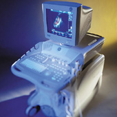 From GE Healthcare.;
(CV Ultrasound) Vivid 3 - 'Vivid family system designed to address customer requests for another level of cardiovascular performance, and offer an even wider range of clinical applications in vascular, pediatrics and the OR. From color flow imaging with super-high frame rates to DICOM connectivity, the new Vivid 3 offers a wealth of technological innovations that enhance image quality, productivity and patient care.' Specifications for this system will be available soon. •
(HPD) Harmonic power Doppler is currently one of the most sensitive techniques for detecting ultrasound contrast agents. HPD works by transmitting multiple pulses toward the object to be imaged and detecting the pulse-to-pulse changes in the received echo signals. Second harmonic bandbass filtering is applied to the received signals to exploit the non-linear behavior of scattering from bubbles (clutter). Harmonic power Doppler operates best at high output levels because of increased contrast destruction, and pulse amplitudes close to the maximum allowed are used much of the time. With a high mechanical index, non-linear propagation of the sound will cause significant harmonic components from tissue, and the contrast agent to tissue ratio will decrease. Also called Harmonic Power Angio. See also Multiple Frame Trigger. Further Reading: Basics:
News & More:
•
Real-time mode has been developed to present motion like a movie of the body's inner workings, showing this information at a high rate. The special real-time transducer uses a larger sound beam than for A, B or M-modes. A linear array transducer with multiple crystal elements displays real-time compound B-mode images with up to 100 images per second. At each scan line, one sound pulse is transmitted and all echoes from the surface to the deepest range are received. Then the ultrasound beam moves on to the next scan line position where pulse transmission and echo recording are repeated. See also Compound B-Mode, Pulse Inversion Doppler, and Frame Averaging. Further Reading: News & More:
•
Contrast agents improve the sensitivity of vascular Doppler ultrasound, for example in cerebrovascular sonography or examinations of deep abdominal vessels. They also enlarge the role of transcranial Doppler. Microbubbles can be used with various modes e.g., color and power Doppler imaging, as well as pulsed-wave Doppler to increase the signal intensity. However, the ultrasound system must be suitable for contrast enhanced technology. Microbubbles usually stay within the vascular space; nevertheless, the contrast enhancement is limited to 2−6 minutes caused by physiologic clearance and bubble destruction. Depended on the application, contrast agents can be administered with a different injection rate e.g., bolus injection, slow injection, or continuous infusion. Stable, homogeneous, and prolonged enhancement can be obtained with perfusion, lasting until the infusion is stopped. See also Cerebrovascular Ultrasonography, Multiple Frame Trigger. • 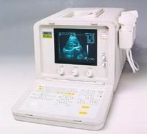 From SIUI Inc.;
'The CTS-385 Plus is designed for the diagnosis of liver, gallbladder, kidney, pancreas, thyroid, breast, uterus, bladder, ovary, etc. The system is a portable linear and convex unit for general application.' Features: 'High quality image Cineloop − 32-frame non-volatile storage capacity Probe frequency conversation option Computer image communication Various measuring function Foldaway keyboard for easy operation Dual probe connector'
Device Information and Specification
APPLICATIONS
See description above
CONFIGURATION
Portable, gray scale(256)
Linear and convex
PROBES STANDARD
1 * 2.5MHz ~ 5.0MHz trifrequency convex probe
2.5MHz to 10.0MHz, linear and convex, broad band, trifrequency
IMAGING OPTIONS
OPTIONAL PACKAGE
DATA PROCESSING
Pre-processing, correlation-processing, interpolation
H*W*D m
0.26 * 0.3 * 0.41
WEIGHT
10 - 13 kg
POWER REQUIREMENT
AC 220V/110V, 50Hz/60Hz
POWER CONSUMPTION
0.1 KVA
Result Pages : | Share This Page Look Ups |
Medical-Ultrasound-Imaging.com
former US-TIP.com
Member of SoftWays' Medical Imaging Group - MR-TIP • Radiology TIP • Medical-Ultrasound-Imaging
Copyright © 2008 - 2024 SoftWays. All rights reserved.
Terms of Use | Privacy Policy | Advertise With Us
former US-TIP.com
Member of SoftWays' Medical Imaging Group - MR-TIP • Radiology TIP • Medical-Ultrasound-Imaging
Copyright © 2008 - 2024 SoftWays. All rights reserved.
Terms of Use | Privacy Policy | Advertise With Us
[last update: 2023-11-06 01:42:00]