Medical Ultrasound Imaging
Sunday, 19 May 2024
Info Sheets Out- side     | 'Linear Array Transducer' p3 Searchterm 'Linear Array Transducer' found in 22 articles 1 term [ • ] - 12 definitions [• ] - 9 booleans [• ]Result Pages : •
Transducers used for the real-time mode are different than for the A-mode, B-, or M-modes. A linear array transducer with multiple piezoelectric crystal elements that are different arranged and fired, transmits the needed larger sound beam. A subgroup of x adjacent elements (8-16; or more in wide-aperture designs) is pulsed simultaneously; the inner elements pulse delayed with respect to the outer elements. The interference of the x small divergent wavelets generates a focused beam. The delay time determining the focus depth of a real-time transducer can be changed during imaging. Similar delay factors applied during the receiving phase, result in a dynamic focusing effect on the return. This forms a single scan line in the real-time image. To produce the following scan line, another group of x elements is selected by shifting one element position along the transducer array from the previous group. This pattern is then repeated for the groups along the array, in a sequential and repetitive way. Further Reading: Basics: •
Transducers can be divided in: 1.) Transducers where the sound wave is transmitted and received by different elements. 2.) Transducers where multiple elements part of the time transmit and part of the time receive sound energy. The first type of ultrasound transducer is used in detection of blood flow (also called nonimaging transducers). For example, the continuous wave transducer (Pedoff transducer) has two separate elements, where one element is always transmitting while the other element is always receiving. Probes of the second type are used to image cardiac structures and have the capability to use various Doppler techniques to detect blood flow (also called imaging transducers). For example, continuous wave, pulsed wave, high pulse repetition frequency, color flow, M-mode, and 2D-mode are the various modes that this type of transducer can perform. Transducers can also be divided in mechanical and electronic or phased scan types. Mechanical transducers use a combination of single element oscillation, multiple element rotation, or a single element and set of acoustic mirrors to generate the sweeping beam for 2D mode. Caused by the vibration (created as the mirrors rotate or oscillate inside the cover) is this type sometimes called the 'wobbler'. Mechanical transducers are cheaper than electronic transducers. Different types of electronic or phased array probes can create a linear or rectangular shaped scan plane as well as a sector or pie shaped scan plane. Sector scanners are most useful for cardiac ultrasound examinations where the beam is directed between the ribs to image the heart. A linear array transducer is more useful in abdominal, OB/GYN, and small parts examinations. Electronic transducers are more expensive but they provide dynamic focusing and smaller probe. See also Rectangular Array Transducer. •
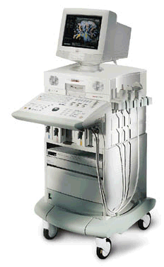
Ultrasound machines, widely used in medical imaging, are essential tools in the field of diagnostic ultrasound. These devices utilize high-frequency sound waves to create real-time images of internal body structures. Ultrasound machines consist of several key components that work together to generate diagnostic images.
These include:
•
The transducer is a handheld device that emits and receives sound waves. It converts electrical energy into sound waves and captures the returning echoes to create images.
•
The control panel houses the interface where the sonographer adjusts imaging parameters such as depth, frequency, and gain. It allows for customization of imaging settings based on the clinical requirements. The transducer pulse controls change the amplitude, frequency and duration of the pulses emitted from the transducer probe.
•
The central processing unit (CPU) serves as the brain of the ultrasound machine, processing the acquired data and transforming it into images. It handles complex calculations, image optimization, data storage and contains the electrical power supplies for itself and the transducer probe.
•
The display monitor (oscilloscope, tablet, computer monitor, etc.) showcases the real-time ultrasound images produced by the machine. It provides visual feedback to the sonographer, aiding in the interpretation and analysis of anatomical structures. Handheld ultrasound devices and mobile ultrasound probes can be connected wirelessly to a smartphone or tablet via Bluetooth or WiFi. These end device serves then as the ultrasound monitor.
•
Data input and measurements are done with the keyboard cursor (trackball). Ultrasound devices used for handheld point of care ultrasound (HPOCUS) are operated via the touch screen of the control panel.
•
Images are captured, reviewed, stored and transmitted digitally, using a standard format for digital imaging and communications in medicine (DICOM). Disk storage devices (FDD, HDD, CD, DVD) are outdated, but may be used in older machines to store the acquired images if no picture archiving and communication system (PACS) connection is possible.
•
The displayed ultrasound pictures are usually digitally stored in a PACS. The images from portable ultrasound machines can be stored and conveniently managed on the end device itself, the inserted memory card or in the cloud. With a QR scanner, the images can be accessed via the Internet in the cloud. Often there is also the possibility to get a picture of a baby sonography as a printout.
B-mode machines represent the vast majority of machines used in echocardiology, obstetrical scans, abdominal scans, gynecological scans, etc. B-mode ultrasound machines usually produce the sector (or pie segment-shaped) scans. These ultrasound scans require either a mechanical scanner transducer (the transducer moves to produce the sector scan), or a linear array transducer operated as a phased array. Ultrasound machines come in different types, each catering to specific clinical needs. The two primary types are stationary and portable ultrasound machines: •
Stationary units are typically larger in size and are installed in dedicated imaging rooms. These machines offer advanced imaging capabilities and a wide range of specialized features. They are commonly found in hospitals, clinics, and university medical centers where comprehensive imaging services are provided.
•
Portable units (see Portable Ultrasound Machine), as the name suggests, are compact and lightweight, designed for on-the-go imaging. These machines are highly versatile and offer excellent mobility, allowing healthcare professionals to bring the ultrasound system directly to the patient's bedside. Portable ultrasound machines are particularly useful in emergency settings, rural healthcare facilities, and point-of-care applications.
See also Handheld Ultrasound, Ultrasound System Performance, Equipment Preparation, Coaxial Cable, and Microbubble Scanner Modification, Environmental Protection and Ultrasound Accessories and Supplies. Further Reading: Basics: News & More:
•  From ESAOTE S.p.A.;
'The AU5 EPI is an ultrasound system for multi-disciplinary use that incorporates all the transducer technologies (linear, convex, phased array, and annular array) adapted to today's most diverse diagnostic requirements. Furthermore it offers the most modern and advanced techniques available today on the market.' • 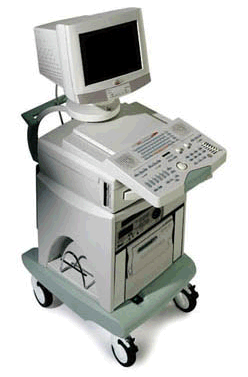 From ESAOTE S.p.A.;
'The AU5 EPI is an ultrasound system for multi-disciplinary use that incorporates all the transducer technologies (linear, convex, phased array, and annular array) adapted to today's most diverse diagnostic requirements. Furthermore it offers the most modern and advanced techniques available today on the market.' Result Pages : | Share This Page Look Ups |
Medical-Ultrasound-Imaging.com
former US-TIP.com
Member of SoftWays' Medical Imaging Group - MR-TIP • Radiology TIP • Medical-Ultrasound-Imaging
Copyright © 2008 - 2024 SoftWays. All rights reserved.
Terms of Use | Privacy Policy | Advertise With Us
former US-TIP.com
Member of SoftWays' Medical Imaging Group - MR-TIP • Radiology TIP • Medical-Ultrasound-Imaging
Copyright © 2008 - 2024 SoftWays. All rights reserved.
Terms of Use | Privacy Policy | Advertise With Us
[last update: 2023-11-06 01:42:00]