Medical Ultrasound Imaging
Wednesday, 8 May 2024
Info Sheets Out- side     | 'Liver Imaging' p2 Searchterm 'Liver Imaging' found in 20 articles 20 booleans [ • ]Result Pages : • 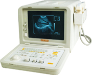 From SIUI Inc.;
'SIUI's newly developed CTS-485 is a portable ultrasound system with advanced technology. With its powerful features, such as 256 grayscale, cineloop, RS232C interface and broadband high-density, multi-frequency probes, the system is designed for professional applications in cardiology, abdominal, small parts, liver, gallbladder, kidney, obstetrics, gynecology, peripheral vascular, etc. The CTS-485 has passed FDA clearance and CE Marking.'
Device Information and Specification
APPLICATIONS
CONFIGURATION
Portable, gray scale(256)
Linear and convex
PROBES STANDARD
1 * 2.5MHz ~ 5.0MHz multifrequency convex probe
2.5MHz to 10.0MHz broad band, trifrequency
IMAGING OPTIONS
Multi zoom rate and depth shift
OPTIONAL PACKAGE
H*W*D m
0.26 * 0.3 * 0.41
WEIGHT
11 kg (main unit)
POWER REQUIREMENT
AC 220V/110V, 50Hz/60Hz
POWER CONSUMPTION
0.1 KVA
•  From SIUI Inc.;
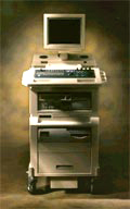
'The Apogee 800Plus ultrasound system offers a new level of high quality in image. With broad clinical application, the system delivers ease of use for cardiac and vascular exams.' 'Due to the exceptional image quality, the sensitivity of color Doppler and many other advanced features, the Apogee 800Plus offers complete diagnosis capability for your entire patient population, including abdominal, obstetrics, gynecology, adult cardiology, pediatric cardiology, peripheral vascular, small parts, breast, prostate, transcranial, etc.'
Device Information and Specification
APPLICATIONS
Linear, curved, phased and symmetric phased array
2D, M-mode, Pulsed wave Doppler, Color Power Angio Imaging, Color 2D, Color M-mode, High PRF Doppler
H*W*D m
1.46 * 0.69 * 0.87
WEIGHT
140 kg (without peripherals)
POWER REQUIREMENT
115Vac, (V~), @ 50 to 60 Hz, 1.2kVA
230Vac, (V~), @ 50 to 60 Hz, 1.2kVA POWER CONSUMPTION
800 Watts (with optional OEM'S: 1200 Watts max)
•  From SIUI Inc.;
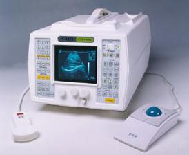
'The CTS-285 is designed for the diagnosis of liver, gallbladder, kidney, pancreas, thyroid, breast, uterus, bladder, ovary, etc. It is a portable versatile ultrasound scanner with convex and linear array scanning.'
Device Information and Specification
APPLICATIONS
See description above
CONFIGURATION
Portable, gray scale(256)
Linear and convex
PROBES STANDARD
1 * 2.5MHz ~ 5.0MHz trifrequency convex probe
2.5MHz to 10.0MHz, linear and convex, broad band, trifrequency
IMAGING OPTIONS
Multi zoom rate and depth shift
DATA PROCESSING
Pre-processing, correlation-processing, interpolation
POWER REQUIREMENT
AC 220V/110V, 50Hz/60Hz
POWER CONSUMPTION
0.06 KVA
•
(LUS) Diagnostic laparoscopy combined with laparoscopic ultrasound is used for staging tumors and to monitor surgical interventions like for example radiofrequency ablation or cryotherapy. Laparoscopic ultrasound provides direct contact imaging of organs with high frequency ultrasound. Laparoscopic ultrasound identifies and characterizes the tumor, guides the probe, and monitors the progression of the freezing or the thermal destruction. This procedure avoid unnecessary open surgery and improves selection of patients for tumor resection e.g., in liver and pancreas. Challenges of LUS are limitations of the intraoperative acoustic windows and the possible movement of the probe and that standard orientation techniques are difficult to apply with laparoscopic instruments, resulting in images from oblique planes. 3D ultrasound or special navigation systems may be helpful. See also Ultrasound Therapy. •
A liver sonography is a diagnostic tool to image the liver and adjoining upper abdominal organs such as the gallbladder, spleen, and pancreas. Deeper structures such as liver and pancreas are imaged at a lower frequency 1-6 MHz with lower axial and lateral resolution but greater penetration. The diagnostic capabilities in this area can be limited by gas in the bowel scattering the sound waves. The application of microbubbles may be useful for detection of liver lesions and for lesion characterization. Some microbubbles have a liver-specific post vascular phase where they appear to be taken up by the reticuloendothelial system (RES). Dynamic contrast enhanced scans in a similar way as with CT or MRI can be used to studying the arterial, venous and tissue phase. After a bolus injection, early vascular enhancement is seen at around 30sec in arterialized lesions (e.g., hepatocellular carcinomas (HCC), focal nodular hyperplasia (FNH)). Later enhancement is typical of hemangiomas with gradually filling towards the center. In the late phase at around 90sec, HCCs appear as defects against the liver background. Most metastases are relatively hypovascular and so do not show much enhancement and are seen as signal voids in the different phases. Either with an intermittent imaging technique or by continuous scanning in a nondestructive, low power mode, characteristic time patterns can be used to differentiate lesions. See also Medical Imaging, B-Mode, High Intensity Focused Ultrasound, Ultrasound Safety and Contrast Medium. Further Reading: Basics:
News & More:
Result Pages : | Share This Page Look Ups |
Medical-Ultrasound-Imaging.com
former US-TIP.com
Member of SoftWays' Medical Imaging Group - MR-TIP • Radiology TIP • Medical-Ultrasound-Imaging
Copyright © 2008 - 2024 SoftWays. All rights reserved.
Terms of Use | Privacy Policy | Advertise With Us
former US-TIP.com
Member of SoftWays' Medical Imaging Group - MR-TIP • Radiology TIP • Medical-Ultrasound-Imaging
Copyright © 2008 - 2024 SoftWays. All rights reserved.
Terms of Use | Privacy Policy | Advertise With Us
[last update: 2023-11-06 01:42:00]