Medical Ultrasound Imaging
Sunday, 12 May 2024
Info Sheets Out- side     | 'Magnet' p4 Searchterm 'Magnet' found in 20 articles 1 term [ • ] - 19 definitions [• ] Result Pages : • 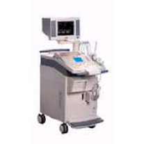 From Siemens Medical Systems;
'The SONOLINE Omnia™ ultrasound system offers mobility, high performance, and ease of use. This digital imaging system delivers excellent 2D, color flow, and Doppler image quality for a variety of general imaging exam types. In addition, the SONOLINE Omnia with cardiac option includes special features that enable use for adult cardiac imaging.'
Device Information and Specification
CLINICAL APPLICATION
Abdomen, small parts, pediatric, prostate, orthopedic, obstetrics, gynecology, cerebrovascular, musculoskeletal, rectal, peripheral vascular (venous and arterial), cardiology
CONFIGURATION
Compact, mobile system
Multi-Frequency and wideband
Wide range of linear/curved/phased array, mechanical, CW pencil probes, laparoscopic, intraoperative, biopsy, TEE transducers
PROBE PORTS
Five
IMAGING OPTIONS
OPTIONAL PACKAGE
Upgradeable applications, cardiac option
IMAGING ENHANCEMENTS
Ultra Fast 3D rendering
STORAGE
Magneto-Optical Drive of 640 MB
DATA PROCESSING
MultiDimensional image processor
• In September 2000 Siemens Medical Engineering Group, bought Acuson Corporation (Mountain View, California) for approximately $700 million. Ultrasound Systems: Ultrasound Systems (older):
Contact Information
•
From Bayer Schering Pharma AG:
Sonovist® (sometimes found as Sonavist) is an investigational ultrasound contrast agent with a biodegradable synthetic capsule filled with sulphur hexafluoride. The biodegradable shell of Sonovist is so stable that it can be taken up by Kupffer cells of the reticuloendothelial system or accumulate in the sinusoids. Therefore, Sonovist® has an additional hepato-splenic parenchymal phase following the blood pool phase, analog to the superparamagnetic iron oxide agents used in liver MRI. The microbubbles are stationary in this phase and generate no conventional Doppler signals. This tissue-specific phase has a variable duration and can be imaged by bubble specific imaging modes.
Drug Information and Specification
RESEARCH NAME
SHU 563A
DEVELOPER
INDICATION
APPLICATION
Intravenous
TYPE
Microbubble
Cyanoacrylate (polymer sheIl)
CHARGE
-
Sulphur hexafluoride
MICROBUBBLE SIZE
-
PRESENTATION
-
STORAGE
-
PREPARATION
-
DO NOT RELY ON THE INFORMATION PROVIDED HERE, THEY ARE
NOT A SUBSTITUTE FOR THE ACCOMPANYING PACKAGE INSERT! •
Ultrasound technology with its advancements is vital for delivering high-quality patient care. Innovations including high-frequency ultrasound, 3D//4D imaging, contrast enhanced ultrasound, elastography, and point-of-care ultrasound, have expanded the capabilities of ultrasound imaging and improved diagnostic accuracy. B-Mode imaging, also known as brightness mode, is the fundamental technique in ultrasound imaging. It produces two-dimensional images based on the echoes received from tissues and organs. Understanding the principles of B-Mode imaging, such as gain adjustment, depth control, and image optimization, is crucial for obtaining diagnostically valuable images. M-Mode imaging, on the other hand, allows for the visualization of motion over time, enabling assessment of cardiac structures and function, as well as fetal heart rate. High-frequency ultrasound refers to the use of ultrasound waves with frequencies greater than 10 MHz. This technology enables improved resolution, allowing for detailed imaging of superficial structures like skin, tendons, and small organs. High-frequency ultrasound has found applications in dermatology, ophthalmology, and musculoskeletal imaging. Traditional 2D ultrasound has been augmented by the advent of 3D ultrasound technology. By acquiring multiple 2D images from different angles, this technique construct a volumetric representation of the imaged area. The addition of 4D ultrasound in real-time motion adds further value by capturing dynamic processes. Doppler imaging employs the Doppler effect to evaluate blood flow within vessels and assess hemodynamics. Color Doppler assigns color to different blood flow velocities, providing a visual representation of blood flow direction and speed. Spectral Doppler displays blood flow velocities as a waveform, allowing for detailed analysis of flow patterns, resistance, and stenosis. Contrast enhanced ultrasound employs microbubble contrast agents to enhance the visualization of blood flow and tissue perfusion. By injecting these agents intravenously, sonographers can differentiate between vascular structures and lesions. Elastography is a technique that measures tissue elasticity or stiffness. It assists in differentiating between normal and abnormal tissues, aiding in the diagnosis of various conditions such as liver fibrosis, breast lesions, and thyroid nodules. Fusion imaging combines ultrasound with other imaging modalities, such as computed tomography (CT), magnetic resonance imaging (MRI), or positron emission tomography (PET). By overlaying or merging ultrasound images with those obtained from other modalities, the user can precisely locate and characterize abnormalities, guide interventions, and improve diagnostic accuracy. Fusion imaging has proven particularly useful in areas such as interventional radiology, oncology, and urology. See also Equipment Preparation, Environmental Protection, Handheld Ultrasound, Portable Ultrasound and Ultrasound Accessories and Supplies. •
Ultrasound therapy uses high energy sound waves to treat different diseases. Historically, the use of ultrasonic waves in therapy began before the wide use as a diagnostic medical imaging tool. Dependend on the intensity, ultrasound therapy reach from the thermal effect used in physical therapy to the destruction of tissue with lithotripsy. Types of ultrasound treatment:
•
See also Thermal Index, History of Ultrasound, Interventional Ultrasound, and B-Mode Acquisition and Targeting. Further Reading: Basics:
News & More:
Result Pages : | Share This Page Look Ups |
Medical-Ultrasound-Imaging.com
former US-TIP.com
Member of SoftWays' Medical Imaging Group - MR-TIP • Radiology TIP • Medical-Ultrasound-Imaging
Copyright © 2008 - 2024 SoftWays. All rights reserved.
Terms of Use | Privacy Policy | Advertise With Us
former US-TIP.com
Member of SoftWays' Medical Imaging Group - MR-TIP • Radiology TIP • Medical-Ultrasound-Imaging
Copyright © 2008 - 2024 SoftWays. All rights reserved.
Terms of Use | Privacy Policy | Advertise With Us
[last update: 2023-11-06 01:42:00]