Medical Ultrasound Imaging
Sunday, 19 May 2024
Info Sheets Out- side     | Ultrasound Database •
(ESWL) Extracorporeal shock wave lithotripsy is a special use of kidney ultrasound, where high intensity focused ultrasound pulses are used to break up calcified stones in the kidney, bladder, or urethra. Pulses of sonic waves pulverize dense renal stones, which are then more easily passed through the ureter and out of the body in the urine. The ultrasound energy at high acoustic power levels is focused to a point exactly on the stone requiring an ultrasound scanning gel for maximum acoustic transmission. Air bubbles in the ultrasound couplant, regardless of their size, degrade the performance of Lithotripsy and have the following effect: Air bubbles smaller that 1/4 wavelength cause scattering of the sound waves as omni directional scatterers and less acoustic energy reaches the focal point. The result is less acoustic power at the focal point to disintegrate the kidney stone. Air bubbles larger than 1/4 wavelength act as reflectors and deflects the acoustic energy off in a different direction. These results in less acoustic energy at the focal point. Microbubbles dispersed throughout the ultrasound couplant layer change the average acoustic impedance of the gel layer (which reduces the total transmitted energy) and, due to refraction, change the focal point. • View NEWS results for 'Lithotripsy' (2). Further Reading: News & More:
•
A liver sonography is a diagnostic tool to image the liver and adjoining upper abdominal organs such as the gallbladder, spleen, and pancreas. Deeper structures such as liver and pancreas are imaged at a lower frequency 1-6 MHz with lower axial and lateral resolution but greater penetration. The diagnostic capabilities in this area can be limited by gas in the bowel scattering the sound waves. The application of microbubbles may be useful for detection of liver lesions and for lesion characterization. Some microbubbles have a liver-specific post vascular phase where they appear to be taken up by the reticuloendothelial system (RES). Dynamic contrast enhanced scans in a similar way as with CT or MRI can be used to studying the arterial, venous and tissue phase. After a bolus injection, early vascular enhancement is seen at around 30sec in arterialized lesions (e.g., hepatocellular carcinomas (HCC), focal nodular hyperplasia (FNH)). Later enhancement is typical of hemangiomas with gradually filling towards the center. In the late phase at around 90sec, HCCs appear as defects against the liver background. Most metastases are relatively hypovascular and so do not show much enhancement and are seen as signal voids in the different phases. Either with an intermittent imaging technique or by continuous scanning in a nondestructive, low power mode, characteristic time patterns can be used to differentiate lesions. See also Medical Imaging, B-Mode, High Intensity Focused Ultrasound, Ultrasound Safety and Contrast Medium. Further Reading: Basics:
News & More:
• •  From GE Healthcare.;
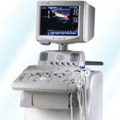
'The new standard in office-based ultrasound.' Specifications for this system will be available soon. • View NEWS results for 'LOGIQ 3' (2). • 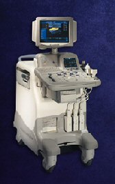 From GE Healthcare.;
Versatile High Performance System 'The LOGIQ 5 Expert ultrasound system delivers the premium performance advantage of TruScan architecture in a versatile high performance package. Advanced capabilities for outstanding clinical performance are available with the efficiency and productivity needed to meet clinical demands.'
Device Information and Specification
APPLICATIONS
Abdominal, cardiac, musculoskeletal, neonatal, OB/GYN, small parts, transcranial, urologic, vascular
CONFIGURATION
Normal system
B-mode, M-mode, anatomic M-mode creation and adjustment, triplex, 3D ultrasound, pulsed wave Doppler, continuous wave Doppler, power Doppler, color Doppler, spectral Doppler
IMAGING OPTIONS
OPTIONAL PACKAGE
STORAGE, CONNECTIVITY, OS
On-board patient, image and reporting archive, HDD, CD-ROM disk burner included, PCMCIA, USB, Windows-based OS
DATA PROCESSING
H*W*D m (inch.)
1.45 * 0.52 * 0.99 (57 * 20 * 39)
WEIGHT
180 kg (397 lbs.)
POWER CONSUMPTION
less than 1.5 KVA
| Share This Page Look Ups |
Medical-Ultrasound-Imaging.com
former US-TIP.com
Member of SoftWays' Medical Imaging Group - MR-TIP • Radiology TIP • Medical-Ultrasound-Imaging
Copyright © 2008 - 2024 SoftWays. All rights reserved.
Terms of Use | Privacy Policy | Advertise With Us
former US-TIP.com
Member of SoftWays' Medical Imaging Group - MR-TIP • Radiology TIP • Medical-Ultrasound-Imaging
Copyright © 2008 - 2024 SoftWays. All rights reserved.
Terms of Use | Privacy Policy | Advertise With Us
[last update: 2023-11-06 01:42:00]