Medical Ultrasound Imaging
Wednesday, 8 May 2024
'3D Ultrasound' p3 Searchterm '3D Ultrasound' found in 23 articles 1 term [ • ] - 18 definitions [• ] - 4 booleans [• ]Result Pages : •  From GE Healthcare.;
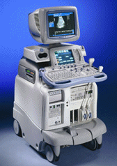
'The System of Choice for General Imaging Imagine a leading-edge ultrasound system so versatile that it can meet the demands of virtually any clinical setting. With the LOGIQ® 9, you'll have a high-performance system capable of multi-dimensional imaging for a full range of clinical applications - from abdominal to breast to vascular imaging. And an ergonomic design that improves scanning comfort and clinical work flow. Now, imagine what LOGIQ® 9 could do for you and your patients.'
Device Information and Specification
APPLICATIONS
Abdominal, cardiac, breast, intraoperative, musculoskeletal, neonatal, OB/GYN, orthopedic, pediatric, small parts, transcranial, urologic, vascular
CONFIGURATION
17' high resolution non-interlaced flat CRT, 4 active probe ports
B-mode, M-mode, coded harmonic imaging, color flow mode (CFM), power Doppler imaging (PDI), PW-HPRF, CW Doppler, color Doppler, pulsed wave Doppler, tissue harmonic imaging
IMAGING OPTIONS
CrossXBeam spatial compounding, coded ultrasound acquisition), speckle reduction imaging (SRI), TruScan technology store raw data, real-time 4D ultrasound, Tru 3D ultrasound
STORAGE, CONNECTIVITY, OS
Patient and image archive, HDD, DICOM 3.0, CD/DVD, MOD, PCMCIA, USB, Windows-based
DATA PROCESSING
Digital beamformer with 1024 system processing channel technology
H*W*D m (inch.)
1.62 * 0.61 * 0.99 (64 * 24 * 39)
WEIGHT
202 kg (408 lb.)
POWER CONSUMPTION
less than 2 KVA
•
(LUS) Diagnostic laparoscopy combined with laparoscopic ultrasound is used for staging tumors and to monitor surgical interventions like for example radiofrequency ablation or cryotherapy. Laparoscopic ultrasound provides direct contact imaging of organs with high frequency ultrasound. Laparoscopic ultrasound identifies and characterizes the tumor, guides the probe, and monitors the progression of the freezing or the thermal destruction. This procedure avoid unnecessary open surgery and improves selection of patients for tumor resection e.g., in liver and pancreas. Challenges of LUS are limitations of the intraoperative acoustic windows and the possible movement of the probe and that standard orientation techniques are difficult to apply with laparoscopic instruments, resulting in images from oblique planes. 3D ultrasound or special navigation systems may be helpful. See also Ultrasound Therapy. •
(MIP) Angiography (Doppler) images can be processed by Maximum Intensity Projection to interactively create different projections. Although the maximum intensity projection (MIP) post processing algorithm is sensitive to high signal from inflowing spins as used in MRI, it is also sensitive to high signal of any other etiology as used in ultrasound imaging. The MIP connects the high intensity dots of the blood vessels in three dimensions, providing an angiogram that can be viewed from any projection. Each point in the MIP represents the highest intensity experienced in that location on any partition within the imaging volume. For complete interpretation the base slices should also be reviewed individually and with multiplanar reconstruction (MPR) software. The MIP can then be displayed in a CINE format or filmed as multiple images acquired from different projections. See also 3D Ultrasound. •
The prostate is a walnut-shaped gland surrounding the beginning of the urethra in front of the rectum and below the bladder. The prostate can become enlarged (particularly in men over age 50) and develop diseases like prostate cancer or inflammation (prostatitis). A large tumor can be felt by a rectal examination. The most effective way of detecting the early signs of prostate cancer is a combination of a prostate-specific antigen (PSA) blood test and a prostate ultrasound examination. An abnormally high level of PSA can indicate prostate cancer or other prostate diseases such as benign prostatic hypertrophy or prostatitis. The transrectal sonography is an important diagnostic ultrasound procedure in determining whether there is any benign enlargement of the prostate or any abnormal nodules. The imaging is performed with a rectal probe, yielding high resolution. High resolution 3D ultrasound provides reliable and accurate determination of the size and the location of cancer. Additionally, ultrasound elastography is a technique in development to improve the specificity and sensitivity of cancer detection. Ultrasound is also used to detect whether cancerous tissue is still only within the prostate or whether it has begun to spread out and to guide a diagnostic biopsy or ultrasound therapy. See also Brachytherapy, and High Intensity Focused Ultrasound. Further Reading: News & More:
•
The elements of a rectangular array transducer (also called matrix transducer) are arranged in a rectangular pattern. Rectangular arrays with unequal rows (e.g. 3, 5, 7) of transducer elements are in real 2D (two-dimensional), but they are termed 1.5D, because the number of rows is much less than the number of columns. Their main advantage is electronic focusing even in the elevation plane (z-plane). The transducers that are termed 2D have an equal number of rows and columns. 2D transducers have the potential to provide real-time 3D ultrasound imaging without moving the transducer. Active matrix array transducers have several elements in the short axis and in addition multiple elements along the long axis. This allows electronic focusing in both axes, resulting in a narrower elevation axis beam width in the near field and far field. Result Pages : |
Medical-Ultrasound-Imaging.com
former US-TIP.com
Member of SoftWays' Medical Imaging Group - MR-TIP • Radiology TIP • Medical-Ultrasound-Imaging
Copyright © 2008 - 2024 SoftWays. All rights reserved.
Terms of Use | Privacy Policy | Advertise With Us
former US-TIP.com
Member of SoftWays' Medical Imaging Group - MR-TIP • Radiology TIP • Medical-Ultrasound-Imaging
Copyright © 2008 - 2024 SoftWays. All rights reserved.
Terms of Use | Privacy Policy | Advertise With Us
[last update: 2023-11-06 01:42:00]


