Medical Ultrasound Imaging
Sunday, 20 September 2026

 Info Sheets  Out- side     | 'Array' p14 Searchterm 'Array' found in 71 articles 9 terms [ • ] - 62 definitions [• ] Result Pages : • 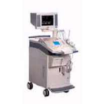 From Siemens Medical Systems;
'The SONOLINE Omnia™ ultrasound system offers mobility, high performance, and ease of use. This digital imaging system delivers excellent 2D, color flow, and Doppler image quality for a variety of general imaging exam types. In addition, the SONOLINE Omnia with cardiac option includes special features that enable use for adult cardiac imaging.'
Device Information and Specification
CLINICAL APPLICATION
Abdomen, small parts, pediatric, prostate, orthopedic, obstetrics, gynecology, cerebrovascular, musculoskeletal, rectal, peripheral vascular (venous and arterial), cardiology
CONFIGURATION
Compact, mobile system
Multi-Frequency and wideband
Wide range of linear/curved/phased array, mechanical, CW pencil probes, laparoscopic, intraoperative, biopsy, TEE transducers
PROBE PORTS
Five
IMAGING OPTIONS
OPTIONAL PACKAGE
Upgradeable applications, cardiac option
IMAGING ENHANCEMENTS
Ultra Fast 3D rendering
STORAGE
Magneto-Optical Drive of 640 MB
DATA PROCESSING
MultiDimensional image processor
•
The dimension of the ultrasound beam and the transducer array are the origin of the slice thickness artifact. This artifact is also called partial volume artifact or volume averaging artifact. See also Validation. •
A tri-frequency probe emits three different frequencies. Probes with tri-frequency capabilities allow a wide range of scanning applications from a single probe. See also Multi-Frequency Probe and Dual Frequency Phased Array Transducer. •
Unlike regular sound, ultrasound can be directed into a single direction. The echoes received by a stationary probe will result in a single dimensional signal showing peaks for every major material change. To generate a 2D picture, the probe is swiveled, either mechanically or through a phased array of ultrasound transducers. The data is analyzed by computer and used to construct the image. In a similar way, 3D pictures can be generated by computer using a specialized probe. In this way, a photo of an unborn baby may be made. Some ultrasonography machines can produce color pictures, of sorts. Doppler ultrasonography is color coded onto a gray scale picture. From the amount of energy in each echo, the difference in acoustic impedance can be calculated and a color is then assigned accordingly. See also Densitometry and 3D Ultrasound. • 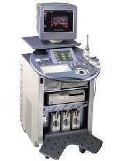 From GE Healthcare.;
'GE is defining a new age of ultrasound. We call it Volume Ultrasound. GE's Voluson 730 Expert is a powerful system that enables real-time techniques for acquiring, navigating and analyzing volumetric images so that you can make clinical decisions with unprecedented confidence.'
Device Information and Specification
APPLICATIONS
Abdominal, breast, cardiac, musculoskeletal, neonatal, OB/GYN, pediatric, small parts, transcranial, urological, vascular
CONFIGURATION
15' high resolution non-interlaced flat CRT, 4 active probe ports
B-mode, M-mode, coded harmonic imaging (2-D), color flow mode (CFM), power Doppler imaging (PDI), color Doppler, pulsed wave Doppler, high pulse repetition frequency (HPRF) Doppler, tissue harmonic imaging, 3-D power Doppler
IMAGING OPTIONS
CrossXBeam spatial compounding, coded excitation , spatio-temporal image correlation (STIC), B-Flow (simultaneous imaging of tissue and blood flow), strain rate imaging (SRI)
OPTIONAL PACKAGE
STORAGE, CONNECTIVITY, OS
SonoView archiving and data management, network, HDD, DICOM 3.0, CD/DVD, MOD, USB, Windows-based
DATA PROCESSING
Digital beamformer with 512 system processing channel technology
H*W*D m (inch.)
1.43 * 0.69 * 1.02 (56 * 27 * 40)
WEIGHT
136 kg (300 lbs.)
Result Pages : | Share This Page     Look Ups |

Medical-Ultrasound-Imaging.com
former US-TIP.com
Member of SoftWays' Medical Imaging Group - MR-TIP • Radiology TIP • Medical-Ultrasound-Imaging
Copyright © 2008 - 2025 SoftWays. All rights reserved.
Terms of Use | Privacy Policy | Advertise With Us
former US-TIP.com
Member of SoftWays' Medical Imaging Group - MR-TIP • Radiology TIP • Medical-Ultrasound-Imaging
Copyright © 2008 - 2025 SoftWays. All rights reserved.
Terms of Use | Privacy Policy | Advertise With Us
[last update: 2025-04-05 02:38:00]