Medical Ultrasound Imaging
Sunday, 20 September 2026

 Info Sheets  Out- side     | 'Resolution' p8 Searchterm 'Resolution' found in 71 articles 5 terms [ • ] - 66 definitions [• ] Result Pages : •
(EUS) Endoscopic ultrasound uses a small probe that is inserted in the rectum either through a proctoscope or by itself. During the test biopsies of any suspicious areas are possible. The usual necessary preparation is an enema to empty the rectum. Endoscopic ultrasound provides additional information about rectal polyps, rectal cancer, perianal infection, and sphincter muscle injuries and improves the selection of patients for local excision. Transrectal echography using a high-frequency transducer is a well established method for preoperative rectal carcinoma assessment. Endoscopic scanning is limited by the ultrasound physics (depth and axial resolution) of the endocavitary probe. Therefore, the combination of endoscopic and transcutaneous ultrasound is most favorable. • 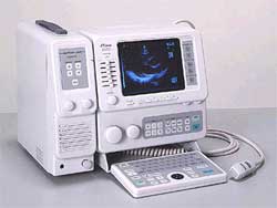 From Fukuda Denshi Co., Ltd.;
'Phased array probes made available for High resolution imaging of full cardiovascular examinations. By adding Linear and Convex probes, the examination areas are extended to Abdominal and OB/GYN applications. B-mode (1, 2 displays), M, and B/M-modes can be individually shown. Optional Doppler unit available for observation of Blood flow information in pulsed and continuous wave Doppler. Portable with IP (Image Processing), Full Keyboard, and track ball are standard.' • 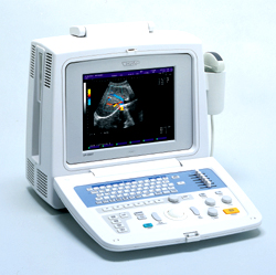 From Fukuda Denshi Co., Ltd.;
'Fully software controlled Image Processing (F-XT technology) Color Doppler & Power Doppler display available Autofocus function for transmission High-resolution SVGA display Cine memory with 128 frames Wide band Probe for multi frequency function 6-step STC adjustment DICOM-compatible Ethernet port 3.5-inch 640MB drive for image filing' •
The focal zone is the region within the transmitted sound beam in which the beam narrows to its minimum size. The lateral resolution is best within the focal zone of the beam.
• 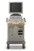 From Philips Medical Systems;
'Clinicians are demanding smaller, higher performing systems specifically designed to meet their clinical and operational challenges. The new Philips HD11 system provides an uncompromising platform, plus advanced options in a highly mobile and easy-to-use system.'
Device Information and Specification
APPLICATIONS
Abdominal, cardiac (also for adults with TEE), musculoskeletal (also pediatric), OB/GYN, prostate, smallparts, transcranial, vascular
CONFIGURATION
17' high resolution non-interlaced flat CRT, 4 active probe ports
B-mode, M-mode, coded harmonic imaging, color flow mode (CFM), power Doppler imaging (PDI), color Doppler, pulsed wave Doppler, tissue harmonic imaging
IMAGING OPTIONS
CrossXBeam spatial compounding, coded ultrasound acquisition),speckle reduction imaging (SRI), TruScan technology store raw data, CINE review with 4 speed types
OPTIONAL PACKAGE
Transesophageal scanning, stress echo, tissue velocity imaging (TVI), tissue velocity Doppler (TVD), contrast harmonic imaging
STORAGE, CONNECTIVITY, OS
Patient and image archive, HDD, DICOM 3.0, CD/DVD, MOD, Windows-based
DATA PROCESSING
Digital beamformer with 1024 system processing channel technology
H*W*D m (inch.)
1.62 * 0.61 * 0.99 (64 * 24 * 39)
WEIGHT
246 kg (498 lbs.)
POWER CONSUMPTION
less than 1.5 KVA
Result Pages : | Share This Page     Look Ups |

Medical-Ultrasound-Imaging.com
former US-TIP.com
Member of SoftWays' Medical Imaging Group - MR-TIP • Radiology TIP • Medical-Ultrasound-Imaging
Copyright © 2008 - 2025 SoftWays. All rights reserved.
Terms of Use | Privacy Policy | Advertise With Us
former US-TIP.com
Member of SoftWays' Medical Imaging Group - MR-TIP • Radiology TIP • Medical-Ultrasound-Imaging
Copyright © 2008 - 2025 SoftWays. All rights reserved.
Terms of Use | Privacy Policy | Advertise With Us
[last update: 2025-04-05 02:38:00]