Medical Ultrasound Imaging
Sunday, 19 May 2024
Info Sheets Out- side     | Ultrasound Database • 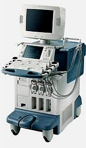 From Toshiba America Medical Systems Inc.;
'Enhanced navigation, visualization, quantification and communication characterize the Aplio ultrasound system, a powerful diagnostic imaging tool and flexible research instrument.' • View NEWS results for 'Toshiba Aplio' (2). •
(TCCS) Transcranial color coded sonography is a combination of B-mode and pulsed wave Doppler. TCCS is used to study morphological and functional assessment of the circle of Willis, intracranial hemodynamics caused by extracranial artery stenosis, collateral flow and the vascular supply of intracranial lesion. Color imaging of the intracranial vessels allows placing the spectral Doppler volume correctly. This modality has encouraged the widespread use. Contrast enhanced TCCS analysis of cerebral arteriovenous transit time (cTT) is used as a measure of cerebral microcirculation. The windows that are used for transcranial Doppler examinations include regions where the skull bones are relatively thin or where naturally occurring gaps allow proper penetration of the sound beam. See also A-Mode, Cranial Bone Thermal Index, Transcranial Doppler and Transcranial Window. •
(TCD) Transcranial color Doppler sonography allows to evaluate the presence and flow direction of vessels as well as their relationships to surrounding structures. A disadvantage of cerebrovascular ultrasonography is the attenuation of the ultrasound signal by the skull. The loss of power through the skull is considerable, the signal to noise ratio is poor and so contrast enhanced Doppler imaging is advantageous. The use of ultrasound contrast agents provides a diagnostic window of sufficient duration and imaging quality to improve an evaluation of the cerebral vessels. Contrast TCD also results in visualization of small arteries and veins and greater length of these vessels. See also A-Mode, Cranial Bone Thermal Index, Transcranial Color Coded Sonography and Transcranial Window. • View NEWS results for 'Transcranial Doppler' (4). Further Reading: Basics:
News & More:
•
The first step in a transcranial Doppler (TCD) examination is to localize a cranial acoustic window where the ultrasound beam can penetrate without being excessively attenuated. There are three main transcranial acoustic windows, used for the sound beam in cerebrovascular ultrasonography to overcome the skull barrier: A complete TCD examination incorporates these windows allowing the visualization of the complete cerebral circulation. Further Reading: News & More:
•
A transducer is a device, usually electrical or electronic, that converts one type of energy to another. Most transducers are either sensors or actuators. A transducer (also called probe) is a main part of the ultrasound machine. The transducer sends ultrasound waves into the body and receives the echoes produced by the waves when it is placed on or over the body part being imaged. Ultrasound transducers are made from crystals with piezoelectric properties. This material vibrates at a resonant frequency, when an alternating electric current is applied. The vibration is transmitted into the tissue in short bursts. The speed of transmission within most soft tissues is 1540 m/s, producing a transit time of 6.5 ms/cm. Because the velocity of ultrasound waves is constant, the time taken for the wave to return to the transducer can be used to determine the depth of the object causing the reflection. The waves will be reflected when they encounter a boundary between two tissues of different density (e.g. soft tissue and bone) and return to the transducer. Conversely, the crystals emit electrical currents when sound or pressure waves hit them (piezoelectric effect). The same crystals can be used to send and receive sound waves; the probe then acts as a receiver, converting mechanical energy back into an electric signal which is used to display an image. A sound absorbing substance eliminates back reflections from the probe itself, and an acoustic lens focuses the emitted sound waves. Then, the received signal gets processed by software to an image which is displayed at a monitor. Transducer heads may contain one or more crystal elements. In multi-element probes, each crystal has its own circuit. The advantage is that the ultrasound beam can be controlled by changing the timing in which each element gets pulsed. Especially for cardiac ultrasound it is important to steer the beam. Usually, several different transducer types are available to select the appropriate one for optimal imaging. Probes are formed in many shapes and sizes. The shape of the probe determines its field of view. Transducers are described in megahertz (MHz) indicating their sound wave frequency. The frequency of emitted sound waves determines how deep the sound beam penetrates and the resolution of the image. Most transducers are only able to emit one frequency because the piezoelectric ceramic or crystals within it have a certain inherent frequency, but multi-frequency probes are also available. See also Blanking Distance, Damping, Maximum Response Axis, Omnidirectional, and Huygens Principle. • View NEWS results for 'Transducer' (17). Further Reading: News & More:
| Share This Page Look Ups |
Medical-Ultrasound-Imaging.com
former US-TIP.com
Member of SoftWays' Medical Imaging Group - MR-TIP • Radiology TIP • Medical-Ultrasound-Imaging
Copyright © 2008 - 2024 SoftWays. All rights reserved.
Terms of Use | Privacy Policy | Advertise With Us
former US-TIP.com
Member of SoftWays' Medical Imaging Group - MR-TIP • Radiology TIP • Medical-Ultrasound-Imaging
Copyright © 2008 - 2024 SoftWays. All rights reserved.
Terms of Use | Privacy Policy | Advertise With Us
[last update: 2023-11-06 01:42:00]