Medical Ultrasound Imaging
Friday, 17 May 2024
Info Sheets Out- side     | 'Center Frequency' Searchterm 'Center Frequency' found in 5 articles 1 term [ • ] - 1 definition [• ] - 3 booleans [• ]Result Pages : • Center Frequency
The center frequency is the median frequency of the transmitted pulse. This pulse contains a range of frequencies in pulsed ultrasound systems.
•
Harmonic imaging relies on detection of harmonics of the transmitted
frequency produced by bubble oscillation. This method is widely available on ultrasound scanners and uses the same array transducers as conventional imaging. A major limitation of the use of ultrasound contrast agents is the problem that signals from the microbubbles are mixed with those from tissue. Echoes from solid tissue and red blood cells are suppressed by harmonic imaging. In harmonic mode, the system transmits at one frequency, but is tuned to receive echoes preferentially at double that frequency, and the second harmonic echoes from the place of the bubble. Typically, the transmit frequency lies between 1.5 and 3 MHz and the receive frequency is selected by means of a bandpass filter whose center frequency lies between 3 and 6 MHz. Color Doppler and real-time harmonic spectral Doppler modes have also been implemented and show a level of tissue motion suppression not available in conventional modes. See also Harmonic B-Mode Imaging, and Harmonic Power Doppler. Further Reading: Basics:
News & More:
•  From Ultralink LLC;
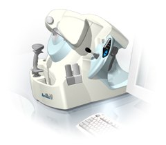
'Artemis is a very high frequency (VHF) ultrasound eye scanner. In use, the patient leans forward placing their head onto an adjustable headrest. The headrest's unique design permits the patient to pull away quickly from the scanner if desired. An eyecup filled with a saline-based interface fluid couples the ultrasound signal to the eye, while a precision mechanism moves the transducer past the front of the eye. During the accurately controlled arc motion of the transducer, which lasts less than one second, many thousands of ultrasound samples are digitized. Following a scan, signal analysis is performed on a PC-compatible microcomputer, and the data are available for immediate viewing on an LCD monitor or disk storage. Artemis is very flexible; many adjustments to the scanning parameters are possible to customize the scan to your clinical needs. Functions are provided for centering the scan about the optical axis of the eye. The starting location of the scans as well as the extent can be varied as desired, to view image planes through the eye at different angles.' See also Ultrasound Biomicroscopy, A-Mode and A-Scan. Further Reading: News & More:
•
A liver sonography is a diagnostic tool to image the liver and adjoining upper abdominal organs such as the gallbladder, spleen, and pancreas. Deeper structures such as liver and pancreas are imaged at a lower frequency 1-6 MHz with lower axial and lateral resolution but greater penetration. The diagnostic capabilities in this area can be limited by gas in the bowel scattering the sound waves. The application of microbubbles may be useful for detection of liver lesions and for lesion characterization. Some microbubbles have a liver-specific post vascular phase where they appear to be taken up by the reticuloendothelial system (RES). Dynamic contrast enhanced scans in a similar way as with CT or MRI can be used to studying the arterial, venous and tissue phase. After a bolus injection, early vascular enhancement is seen at around 30sec in arterialized lesions (e.g., hepatocellular carcinomas (HCC), focal nodular hyperplasia (FNH)). Later enhancement is typical of hemangiomas with gradually filling towards the center. In the late phase at around 90sec, HCCs appear as defects against the liver background. Most metastases are relatively hypovascular and so do not show much enhancement and are seen as signal voids in the different phases. Either with an intermittent imaging technique or by continuous scanning in a nondestructive, low power mode, characteristic time patterns can be used to differentiate lesions. See also Medical Imaging, B-Mode, High Intensity Focused Ultrasound, Ultrasound Safety and Contrast Medium. Further Reading: Basics:
News & More:
•
Ultrasound machines, widely used in medical imaging, are essential tools in the field of diagnostic ultrasound. These devices utilize high-frequency sound waves to create real-time images of internal body structures. Ultrasound machines consist of several key components that work together to generate diagnostic images.
These include:
•
The transducer is a handheld device that emits and receives sound waves. It converts electrical energy into sound waves and captures the returning echoes to create images.
•
The control panel houses the interface where the sonographer adjusts imaging parameters such as depth, frequency, and gain. It allows for customization of imaging settings based on the clinical requirements. The transducer pulse controls change the amplitude, frequency and duration of the pulses emitted from the transducer probe.
•
The central processing unit (CPU) serves as the brain of the ultrasound machine, processing the acquired data and transforming it into images. It handles complex calculations, image optimization, data storage and contains the electrical power supplies for itself and the transducer probe.
•
The display monitor (oscilloscope, tablet, computer monitor, etc.) showcases the real-time ultrasound images produced by the machine. It provides visual feedback to the sonographer, aiding in the interpretation and analysis of anatomical structures. Handheld ultrasound devices and mobile ultrasound probes can be connected wirelessly to a smartphone or tablet via Bluetooth or WiFi. These end device serves then as the ultrasound monitor.
•
Data input and measurements are done with the keyboard cursor (trackball). Ultrasound devices used for handheld point of care ultrasound (HPOCUS) are operated via the touch screen of the control panel.
•
Images are captured, reviewed, stored and transmitted digitally, using a standard format for digital imaging and communications in medicine (DICOM). Disk storage devices (FDD, HDD, CD, DVD) are outdated, but may be used in older machines to store the acquired images if no picture archiving and communication system (PACS) connection is possible.
•
The displayed ultrasound pictures are usually digitally stored in a PACS. The images from portable ultrasound machines can be stored and conveniently managed on the end device itself, the inserted memory card or in the cloud. With a QR scanner, the images can be accessed via the Internet in the cloud. Often there is also the possibility to get a picture of a baby sonography as a printout.
B-mode machines represent the vast majority of machines used in echocardiology, obstetrical scans, abdominal scans, gynecological scans, etc. B-mode ultrasound machines usually produce the sector (or pie segment-shaped) scans. These ultrasound scans require either a mechanical scanner transducer (the transducer moves to produce the sector scan), or a linear array transducer operated as a phased array. Ultrasound machines come in different types, each catering to specific clinical needs. The two primary types are stationary and portable ultrasound machines: •
Stationary units are typically larger in size and are installed in dedicated imaging rooms. These machines offer advanced imaging capabilities and a wide range of specialized features. They are commonly found in hospitals, clinics, and university medical centers where comprehensive imaging services are provided.
•
Portable units (see Portable Ultrasound Machine), as the name suggests, are compact and lightweight, designed for on-the-go imaging. These machines are highly versatile and offer excellent mobility, allowing healthcare professionals to bring the ultrasound system directly to the patient's bedside. Portable ultrasound machines are particularly useful in emergency settings, rural healthcare facilities, and point-of-care applications.
See also Handheld Ultrasound, Ultrasound System Performance, Equipment Preparation, Coaxial Cable, and Microbubble Scanner Modification, Environmental Protection and Ultrasound Accessories and Supplies. Further Reading: Basics: News & More:
Result Pages : | Share This Page Look Ups |
Medical-Ultrasound-Imaging.com
former US-TIP.com
Member of SoftWays' Medical Imaging Group - MR-TIP • Radiology TIP • Medical-Ultrasound-Imaging
Copyright © 2008 - 2024 SoftWays. All rights reserved.
Terms of Use | Privacy Policy | Advertise With Us
former US-TIP.com
Member of SoftWays' Medical Imaging Group - MR-TIP • Radiology TIP • Medical-Ultrasound-Imaging
Copyright © 2008 - 2024 SoftWays. All rights reserved.
Terms of Use | Privacy Policy | Advertise With Us
[last update: 2023-11-06 01:42:00]