Medical Ultrasound Imaging
Sunday, 19 May 2024
Info Sheets Out- side     | 'Color Doppler Imaging' p2 Searchterm 'Color Doppler Imaging' found in 23 articles 1 term [ • ] - 5 definitions [• ] - 17 booleans [• ]Result Pages : •
Sonazoid™ is an ultrasound contrast agent (UCA) consisting of stabilized gas microbubbles in an aqueous suspension. Sonazoid™ has overcome the stability problems of first generation USCA and can produce myocardial perfusion images. Myocardial imaging using ultrasound contrast agents provides diagnosis of chronic heart disease and assessment of the coronary arteries and of the coronary blood flow reserve.
Sonazoid™ is taken up by healthy Kupffer cells in the liver and spleen, but break down in high amplitude ultrasound imaging modes such as color Doppler imaging. The bubble rupture produces a transient pressure wave, which results in a characteristic mosaic color pattern from tissues containing the microbubbles (induced acoustic emission). Liver tumors without Kupffer cells will not display the mosaic pattern and can therefore be identified easily.
Drug Information and Specification
RESEARCH NAME
NC100100
DEVELOPER
INDICATION -
DEVELOPMENT STAGE Development in USA and EU suspended
APPLICATION
-
TYPE
Microbubble
Lipid Stabilized (not disclosed)
CHARGE
Negative
Perfluorobutane
MICROBUBBLE SIZE
-
PRESENTATION
-
STORAGE
-
PREPARATION
Reconstitute with 2mL water
DO NOT RELY ON THE INFORMATION PROVIDED HERE, THEY ARE
NOT A SUBSTITUTE FOR THE ACCOMPANYING PACKAGE INSERT! •
(CDFI) Color [colour, Brit.] Doppler flow imaging is a method based on pulsed ultrasound Doppler technology for visualizing direction and velocity of blood flow within the cardiac chambers or blood vessels.
See also Autocorrelation. • 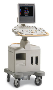 From Philips Medical Systems;
Introduced in June 2005, 'one of the less expensive and more dedicated' ultrasound systems.
Device Information and Specification
CONFIGURATION
LCD monitor
Broadband, convex, linear,
digital beamformer and focal tuning IMAGING OPTIONS
OPTIONAL PACKAGE
DICOM, etc.
STORAGE, CONNECTIVITY, OS
HDD, CD, USB, optionalMOD and DICOM 3.0
DATA PROCESSING
256-digitally processed channels
H*W*D inch.
58 * 20 * 32
WEIGHT
135 lbs.
• 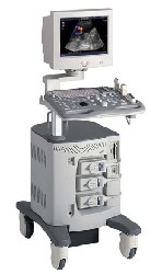 From ALOKA Co., Ltd.;
'A Platform for Digital, Pure-Beam Imaging The high-performance, ALOKA ProSound SSD-3500 utilizes advanced ProSound technologies including: Fully digital beam former A wide dynamic range, 12-bit A/D converter Multi beam processing. The SSD-3500 also helps you achieve more efficient examinations. Its ergonomic, user-friendly design enables you to customize the system according to your specific application needs.'
Device Information and Specification
APPLICATIONS
CONFIGURATION
Compact, portable, dual dynamic display
Color Flow, Power Flow, Spectral Doppler, Real-time Free Angular M-Mode, Tissue Harmonic Imaging, Quint Frequency Imaging, Pure Harmonic Detection
STORAGE, CONNECTIVITY, OS
Data Management Subsystem (iDMS), DICOM-Worklist
DATA PROCESSING
12-bit analog to digital converter
•  From GE Healthcare.;
'GE is defining a new age of ultrasound. We call it Volume Ultrasound. GE's Voluson 730 Expert is a powerful system that enables real-time techniques for acquiring, navigating and analyzing volumetric images so that you can make clinical decisions with unprecedented confidence.'
Device Information and Specification
APPLICATIONS
Abdominal, breast, cardiac, musculoskeletal, neonatal, OB/GYN, pediatric, small parts, transcranial, urological, vascular
CONFIGURATION
15' high resolution non-interlaced flat CRT, 4 active probe ports
B-mode, M-mode, coded harmonic imaging (2-D), color flow mode (CFM), power Doppler imaging (PDI), color Doppler, pulsed wave Doppler, high pulse repetition frequency (HPRF) Doppler, tissue harmonic imaging, 3-D power Doppler
IMAGING OPTIONS
CrossXBeam spatial compounding, coded excitation , spatio-temporal image correlation (STIC), B-Flow (simultaneous imaging of tissue and blood flow), strain rate imaging (SRI)
OPTIONAL PACKAGE
STORAGE, CONNECTIVITY, OS
SonoView archiving and data management, network, HDD, DICOM 3.0, CD/DVD, MOD, USB, Windows-based
DATA PROCESSING
Digital beamformer with 512 system processing channel technology
H*W*D m (inch.)
1.43 * 0.69 * 1.02 (56 * 27 * 40)
WEIGHT
136 kg (300 lbs.)
Result Pages : | Share This Page Look Ups |
Medical-Ultrasound-Imaging.com
former US-TIP.com
Member of SoftWays' Medical Imaging Group - MR-TIP • Radiology TIP • Medical-Ultrasound-Imaging
Copyright © 2008 - 2024 SoftWays. All rights reserved.
Terms of Use | Privacy Policy | Advertise With Us
former US-TIP.com
Member of SoftWays' Medical Imaging Group - MR-TIP • Radiology TIP • Medical-Ultrasound-Imaging
Copyright © 2008 - 2024 SoftWays. All rights reserved.
Terms of Use | Privacy Policy | Advertise With Us
[last update: 2023-11-06 01:42:00]