Medical Ultrasound Imaging
Wednesday, 8 May 2024
'Doppler Spectrum' Searchterm 'Doppler Spectrum' found in 7 articles 1 term [ • ] - 2 definitions [• ] - 4 booleans [• ]Result Pages : • Doppler Spectrum
The Doppler spectrum indicates how the echo power is distributed according to the Doppler shift frequency. The Doppler shift frequency is directly related to the radial velocity of the scatterer.
See also Modal Velocity, Doppler Effect and Doppler Ultrasound. Further Reading: News & More: •
The diastole is the period of relaxation in the cardiac cycle which alternates with systole. During diastole, the ventricles fill and the aortic and pulmonary valves are closed. On a Doppler spectrum analysis, diastole can be identified as beginning at the dicrotic notch (a small abrupt upswing in the deceleration phase of systole) and ending with the systolic upstroke. See also Echocardiography and Cardiac Ultrasound. •
The modal velocity is the frequency component which contains the most energy. In the display of the Doppler spectrum, the mode corresponds to the brightest parts of the individual spectra.
•  From SIUI Inc.;
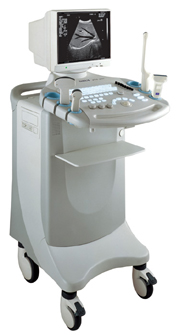
'Dedicated to ultrasound industry, Shantou Institute of Ultrasonic Instruments, Inc. (SIUI) has launched Apogee 3500, the Digital Color Doppler Ultrasound Imaging System. With latest imaging technologies, high-definition image quality and excellent practical functions, the Apogee 3500 offers optimal solutions for clinical ultrasonic examination.' 'The Apogee 3500 is available with many high-density, super broadband and multi-frequency probes, such as convex, micro-convex, linear, vaginal, rectal and phased array probes, which are widely applied for different clinical diagnoses, including abdomen (liver, kidney, gall-bladder, pancreas), gynecology (uterus, ovary), obstetrics (early pregnancy, basic OB, complete OB, multi gestation, fetal echo), cardiology (adult and pediatric cardiology), small parts (thyroid, galactophore, testicles, neonate), peripheral vascular and prostate.'
Device Information and Specification
CONFIGURATION
Normal system, color - gray scale(256)
Linear, convex and phased array
PROBES STANDARD
2.0 MHz ~ 12.0 MHz, broad band, tri-frequency
B-mode (B, 2B, 4B), M-mode, B/M-mode, real-time compound imaging, panoramic imaging, trapezoidal imaging (linear probes), spectrum Doppler (PWD and CWD), color Doppler flow imaging (CDFI), color power angio (CPA), tissue harmonic imaging (THI)
IMAGING OPTIONS
Real-time ZOOM, zoom rate and position selectable
OPTIONAL PACKAGE
H*W*D m
1.29 * 0.52 * 0.75
WEIGHT
110 kg
POWER REQUIREMENT
AC 220V/110V, 50Hz/60Hz
POWER CONSUMPTION
0.6 KVA
•
Spectral analysis is the quantitative analysis method to display the distribution of frequencies. A difficult Doppler signal is separated into the frequency components so that the range of frequencies in a Doppler shifted signal can be analyzed. This allows measurement of blood flow velocity by positioning of a probing cursor in the artery (on the screen), and the signal representing blood flow velocity is generated. The peaks and ebbs create the spectrum, corresponding to systolic and diastolic blood flow. The signal is both visual and auditory.
Further Reading: News & More:
Result Pages : |
Medical-Ultrasound-Imaging.com
former US-TIP.com
Member of SoftWays' Medical Imaging Group - MR-TIP • Radiology TIP • Medical-Ultrasound-Imaging
Copyright © 2008 - 2024 SoftWays. All rights reserved.
Terms of Use | Privacy Policy | Advertise With Us
former US-TIP.com
Member of SoftWays' Medical Imaging Group - MR-TIP • Radiology TIP • Medical-Ultrasound-Imaging
Copyright © 2008 - 2024 SoftWays. All rights reserved.
Terms of Use | Privacy Policy | Advertise With Us
[last update: 2023-11-06 01:42:00]


