Medical Ultrasound Imaging
Wednesday, 8 May 2024
'Gating' p2 Searchterm 'Gating' found in 9 articles 1 term [ • ] - 8 definitions [• ] Result Pages : •
Pressure is the force per unit area applied on a surface in a direction perpendicular to that surface. Pressure can also be described as a form of potential energy in a fluid. The maximum pressure of the fluid medium obtained during propagation of an ultrasonic pulse. The negative peak pressure is the peak rarefaction pressure attained during the negative portion of a propagating ultrasound pulse in a medium such as tissue. Sound pressure can be measured using a microphone in air and a hydrophone in water. The SI unit for sound pressure is the Pascal. Blood pressure is the pressure exerted by the blood on the walls of the blood vessels. See also Rarefactional Pressure, Low Intensity Pulsed Ultrasound, and Projector. •
The Receiver is the component of the ultrasound machine that receives the current generated in the transducer from the returning sound waves. See also Blanking Distance, and Range Gating. •
Sound waves must have a medium to pass through. The velocity or propagation speed is the speed at which sound waves travel through a particular medium measured in meters per second (m/s) or millimeters per microsecond (mm/μs). Because the velocity of ultrasound waves is constant, the time taken for the wave to return to the probe can be used to determine the depth of the object causing the reflection. The velocity is equal to the frequency x wavelength. V = f x l The velocity of ultrasound will differ with different media. In general, the propagation speed of sound through gases is low, liquids higher and solids highest. The speed of sound depends strongly on temperature as well as the medium through which sound waves are propagating. At 0 °C (32 °F) the speed of sound in air is about 331 m/s (1,086 ft/s; 1,192 km/h; 740 mph; 643 kn), at 20 °C (68 °F) about 343 metres per second (1,125 ft/s; 1,235 km/h; 767 mph; 667 kn) Velocity (m/s)
•
air: 331;
•
fat: 1450;
•
water (50 °C): 1540;
•
human soft tissue: 1540;
•
brain: 1541;
•
liver: 1549;
•
kidney: 1561;
•
blood: 1570;
•
muscle: 1585;
•
lens of eye: 1620;
•
bone: 4080.
Doppler ultrasound visualizes blood flow-velocity information. The peak systolic velocity and the end diastolic velocity are major Doppler parameters, which are determined from the spectrum obtained at the point of maximal vessel narrowing. Peak systolic velocity ratios are calculated by dividing the peak-systolic velocity measured at the site of flow disturbance by that measured proximal of the narrowing (stenosis, graft, etc.). See Acceleration Index, Acceleration Time, Modal Velocity, Run-time Artifact and Maximum Velocity. Further Reading: Basics:
News & More:
•  From GE Healthcare.;
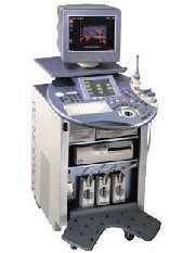
'GE is defining a new age of ultrasound. We call it Volume Ultrasound. GE's Voluson 730 Expert is a powerful system that enables real-time techniques for acquiring, navigating and analyzing volumetric images so that you can make clinical decisions with unprecedented confidence.'
Device Information and Specification
APPLICATIONS
Abdominal, breast, cardiac, musculoskeletal, neonatal, OB/GYN, pediatric, small parts, transcranial, urological, vascular
CONFIGURATION
15' high resolution non-interlaced flat CRT, 4 active probe ports
B-mode, M-mode, coded harmonic imaging (2-D), color flow mode (CFM), power Doppler imaging (PDI), color Doppler, pulsed wave Doppler, high pulse repetition frequency (HPRF) Doppler, tissue harmonic imaging, 3-D power Doppler
IMAGING OPTIONS
CrossXBeam spatial compounding, coded excitation , spatio-temporal image correlation (STIC), B-Flow (simultaneous imaging of tissue and blood flow), strain rate imaging (SRI)
OPTIONAL PACKAGE
STORAGE, CONNECTIVITY, OS
SonoView archiving and data management, network, HDD, DICOM 3.0, CD/DVD, MOD, USB, Windows-based
DATA PROCESSING
Digital beamformer with 512 system processing channel technology
H*W*D m (inch.)
1.43 * 0.69 * 1.02 (56 * 27 * 40)
WEIGHT
136 kg (300 lbs.)
Result Pages : |
Medical-Ultrasound-Imaging.com
former US-TIP.com
Member of SoftWays' Medical Imaging Group - MR-TIP • Radiology TIP • Medical-Ultrasound-Imaging
Copyright © 2008 - 2024 SoftWays. All rights reserved.
Terms of Use | Privacy Policy | Advertise With Us
former US-TIP.com
Member of SoftWays' Medical Imaging Group - MR-TIP • Radiology TIP • Medical-Ultrasound-Imaging
Copyright © 2008 - 2024 SoftWays. All rights reserved.
Terms of Use | Privacy Policy | Advertise With Us
[last update: 2023-11-06 01:42:00]


