Medical Ultrasound Imaging
Saturday, 18 May 2024
Info Sheets Out- side     | 'Pregnancy' p2 Searchterm 'Pregnancy' found in 19 articles 2 terms [ • ] - 17 definitions [• ] Result Pages : •
A-mode (Amplitude-mode) ultrasound is a technique used to assess organ dimensions and determine the depth of an organ. While A-mode technology was previously employed in midline echoencephalography for rapid screening of intracranial mass lesions and ophthalmologic scanning, it is now considered obsolete in medical imaging. Nonetheless, the A-mode scan has found applications in early pregnancy assessment (specifically the detection of fetal heartbeats), cephalometry, and placental localization.
When the ultrasound beam encounters an anatomic boundary, the received sound impulse is processed to appear as a vertical reflection of a point. On the display, it looks like spikes of different heights (the amplitude). The intensity of the returning impulse determined the height of the vertical reflection and the time it took for the impulse to make the round trip would determine the space between verticals. The distance between these spikes can be measured accurately by dividing the speed of sound in tissue (1540 m/sec) by half the sound travel time. During an echoencephalography scan, the first A-mode scan is acquired from the right side of the head and captured on film. Subsequently, the probe is positioned at the corresponding point on the left side, and a second exposure is captured on the same film, displaying inverted spikes. The A-mode ultrasound could be used to identify structures normally located in the midline of the brain such as the third ventricle and falx cerebri. The midline structures would be aligned in normal patients but show displacement in patients with mass lesion such as a subdural, epidural, or intracranial hemorrhage. See also 2D Ultrasound, 3D Ultrasound, 4D Ultrasound, Ultrasound Biomicroscopy, A-scan, B-mode and the Infosheet about ultrasound modes. •
A-scans are used in ophthalmologic scanning, to detect and monitor pregnancy problems, and screen intracranial mass lesions by using A-modes. A-scan ultrasound biometry, commonly referred to as an A-scan, is a routine diagnostic test used in ophthalmology. The A-scan provides data on the shape of the eye, which is a major determinant in common sight disorders. Ultrasound scanners used in this type of test require usually direct contact with the eye. See also A-Mode, Oculoplethysmography, Ultrasound Biomicroscopy, B-Scan, C-Scan and D-Scan. Further Reading: News & More:
•
(AUS) Abdominal ultrasound, also known as abdominal sonography, is a medical imaging technique that focuses on the visualization and assessment of the abdominal organs. While 'abdominal ultrasound' is the commonly used term, there are alternative terms that can be used to refer to this imaging modality: (TAE) transabdominal echography, abdominal ultrasonography, sonogram, FAST (Focused Assessment with Sonography for Trauma). Abdominal ultrasound imaging is an invaluable clinical tool for identifying the underlying cause of abdominal pain. An abdominal ultrasound examination encompasses a comprehensive evaluation of the liver, gallbladder, biliary tree, pancreas, spleen, kidneys, and abdominal blood vessels. It is a cost-effective, safe, and non-invasive medical imaging modality that is typically utilized as the initial diagnostic investigation. Advanced ultrasound techniques, such as high-resolution ultrasound, endoscopic ultrasound, and contrast-enhanced Doppler, further enhance the detection of small lesions and provide detailed information for precise diagnosis. To prepare for an abdominal ultrasound, it is recommended to have nothing to eat or drink for at least 8 hours, starting from midnight the night before the examination. Indications:
•
Abdominal pain
•
Gallbladder or kidneys stones
•
Inflammation
•
Detection of cancer and metastasis
FAST (Focused Assessment with Sonography for Trauma) is a rapid diagnostic test used for trauma patients. It sequentially evaluates the presence of free fluid in the pericardium (hemopericardium) and in four specific views of the abdomen. These views include the right upper quadrant (RUQ), left upper quadrant (LUQ), subcostal, and suprapubic views. They aid in identifying hemoperitoneum in patients with potential truncal injuries. The space between the liver and the right kidney (RUQ), known as Morison's pouch, is a location where intraperitoneal fluid can accumulate. Emergency abdominal ultrasonography is indicated in cases of suspected aortic aneurysm, appendicitis, biliary and renal colic, as well as blunt or penetrating abdominal trauma. It plays a crucial role in the timely assessment and management of these conditions, providing critical information to guide appropriate treatment decisions. See also Handheld Ultrasound, Pelvic Ultrasound, Pregnancy Ultrasound, Prostate Ultrasound, Interventional Ultrasound and Pediatric Ultrasound. Further Reading: Basics: News & More:
•  From SIUI Inc.;
'Dedicated to ultrasound industry, Shantou Institute of Ultrasonic Instruments, Inc. (SIUI) has launched Apogee 3500, the Digital Color Doppler Ultrasound Imaging System. With latest imaging technologies, high-definition image quality and excellent practical functions, the Apogee 3500 offers optimal solutions for clinical ultrasonic examination.' 'The Apogee 3500 is available with many high-density, super broadband and multi-frequency probes, such as convex, micro-convex, linear, vaginal, rectal and phased array probes, which are widely applied for different clinical diagnoses, including abdomen (liver, kidney, gall-bladder, pancreas), gynecology (uterus, ovary), obstetrics (early pregnancy, basic OB, complete OB, multi gestation, fetal echo), cardiology (adult and pediatric cardiology), small parts (thyroid, galactophore, testicles, neonate), peripheral vascular and prostate.'
Device Information and Specification
CONFIGURATION
Normal system, color - gray scale(256)
Linear, convex and phased array
PROBES STANDARD
2.0 MHz ~ 12.0 MHz, broad band, tri-frequency
B-mode (B, 2B, 4B), M-mode, B/M-mode, real-time compound imaging, panoramic imaging, trapezoidal imaging (linear probes), spectrum Doppler (PWD and CWD), color Doppler flow imaging (CDFI), color power angio (CPA), tissue harmonic imaging (THI)
IMAGING OPTIONS
Real-time ZOOM, zoom rate and position selectable
OPTIONAL PACKAGE
H*W*D m
1.29 * 0.52 * 0.75
WEIGHT
110 kg
POWER REQUIREMENT
AC 220V/110V, 50Hz/60Hz
POWER CONSUMPTION
0.6 KVA
• 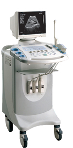
From SIUI Inc.;
Device Information and Specification
CONFIGURATION
Normal system, gray scale(256)
Linear and convex
PROBES STANDARD
2.5MHz ~ 11.0MHz, broad band, tri-frequency
IMAGING OPTIONS
Real zoom, max. Zoom * 4.0, multiplying power and position selectable
OPTIONAL PACKAGE
H*W*D m
1.30 * 0.52 * 0.77
WEIGHT
90kg (main unit)
POWER REQUIREMENT
AC 220V/110V, 50Hz/60Hz
POWER CONSUMPTION
0.45 KVA
Result Pages : | Share This Page Look Ups |
Medical-Ultrasound-Imaging.com
former US-TIP.com
Member of SoftWays' Medical Imaging Group - MR-TIP • Radiology TIP • Medical-Ultrasound-Imaging
Copyright © 2008 - 2024 SoftWays. All rights reserved.
Terms of Use | Privacy Policy | Advertise With Us
former US-TIP.com
Member of SoftWays' Medical Imaging Group - MR-TIP • Radiology TIP • Medical-Ultrasound-Imaging
Copyright © 2008 - 2024 SoftWays. All rights reserved.
Terms of Use | Privacy Policy | Advertise With Us
[last update: 2023-11-06 01:42:00]