Medical Ultrasound Imaging
Sunday, 19 May 2024
Info Sheets Out- side     | 'Range Resolution' p2 Searchterm 'Range Resolution' found in 19 articles 1 term [ • ] - 1 definition [• ] - 17 booleans [• ]Result Pages : •  From Medison Co.,Ltd.;
'Medison's MySono 201 brings the power and flexibility of digital imaging within the reach of everyone. Its lightweight and compact design, featuring 2D black-and-white imaging, a generous 640x480 resolution LCD display, and an extensive range of multifrequency transducers, offers versatility as well as high performance. An AC/DC power supply, outputs for connection to an external monitor, and attractive pricing make the MySono 201 perfect for private practitioners, smaller clinics, and educational facilities.' • 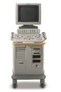 From Philips Medical Systems;
'Clinicians are demanding smaller, higher performing systems specifically designed to meet their clinical and operational challenges. The new Philips HD11 system provides an uncompromising platform, plus advanced options in a highly mobile and easy-to-use system.'
Device Information and Specification
APPLICATIONS
Abdominal, cardiac (also for adults with TEE), musculoskeletal (also pediatric), OB/GYN, prostate, smallparts, transcranial, vascular
CONFIGURATION
17' high resolution non-interlaced flat CRT, 4 active probe ports
B-mode, M-mode, coded harmonic imaging, color flow mode (CFM), power Doppler imaging (PDI), color Doppler, pulsed wave Doppler, tissue harmonic imaging
IMAGING OPTIONS
CrossXBeam spatial compounding, coded ultrasound acquisition),speckle reduction imaging (SRI), TruScan technology store raw data, CINE review with 4 speed types
OPTIONAL PACKAGE
Transesophageal scanning, stress echo, tissue velocity imaging (TVI), tissue velocity Doppler (TVD), contrast harmonic imaging
STORAGE, CONNECTIVITY, OS
Patient and image archive, HDD, DICOM 3.0, CD/DVD, MOD, Windows-based
DATA PROCESSING
Digital beamformer with 1024 system processing channel technology
H*W*D m (inch.)
1.62 * 0.61 * 0.99 (64 * 24 * 39)
WEIGHT
246 kg (498 lbs.)
POWER CONSUMPTION
less than 1.5 KVA
•  From Philips Medical Systems;
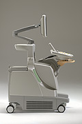
'The Philips iU22 system combines Intelligent Design, including breakthroughs in ergonomics, with Intelligent Control, providing new levels of automation, to give you revolutionary performance and workflow.'
Device Information and Specification
APPLICATIONS
Abdominal, cardiac (also for adults with TEE), musculoskeletal (also pediatric), OB/GYN, prostate, smallparts, transcranial, vascular
CONFIGURATION
17' high resolution non-interlaced flat CRT, 4 active probe ports
B-mode, M-mode, coded harmonic imaging, color flow mode (CFM), power Doppler imaging (PDI), color Doppler, pulsed wave Doppler, tissue harmonic imaging
IMAGING OPTIONS
CrossXBeam spatial compounding, coded ultrasound acquisition),speckle reduction imaging (SRI), TruScan technology store raw data, CINE review with 4 speed types
OPTIONAL PACKAGE
Transesophageal scanning, stress echo, tissue velocity imaging (TVI), tissue velocity Doppler (TVD), contrast harmonic imaging
STORAGE, CONNECTIVITY, OS
Patient and image archive, HDD, DICOM 3.0, CD/DVD, MOD, Windows-based
DATA PROCESSING
Digital beamformer with 1024 system processing channel technology
H*W*D m (inch.)
1.62 * 0.61 * 0.99 (64 * 22 * 43)
WEIGHT
kg (345 lbs.)
POWER CONSUMPTION
•
(PII) Pulse inversion imaging (also called phase inversion imaging) is a non-linear imaging method specifically made for enhanced detection of microbubble ultrasound contrast agents. In PII, two pulses are sent in rapid succession into the tissue; the second pulse is a mirror image of the first. The resulting echoes are added at reception. Linear scattering of the two pulses will give two echoes which are inverted copies of each other, and these echoes will therefore cancel out when added. Linear scattering dominates in tissues. Echoes from linear scatterers such as tissue cancel, whereas those from gas microbubbles do not. Non-linear scattering of the two pulses will give two echoes which do not cancel out completely due to different bubble response to positive and negative pressures of equal magnitude. The harmonic components add, and the signal intensity difference between non-linear and linear scatterers is therefore increased. The resulting images show high sensitivity to bubbles at the resolution of a conventional image. In harmonic imaging, the frequency range of the transmitted pulse and the received signal should not overlap, but this restriction is less in pulse inversion imaging since the transmit frequencies are not filtered out, but rather subtracted. Broader transmit and receive bandwidths are therefore allowed, giving shorter pulses and improved axial resolution, hence the alternative term wideband harmonic imaging. Many ultrasound machines offer some form of pulse inversion imaging. See also Pulse Inversion Doppler, Narrow Bandwidth, Dead Zone, Ultrasound Phantom. •
A matrix is a set of numbers arranged in a rectangular array. The array of numbers is divided in rows and columns. The matrix size determines the scan resolution.
Further Reading: Basics:
News & More:
Result Pages : | Share This Page Look Ups |
Medical-Ultrasound-Imaging.com
former US-TIP.com
Member of SoftWays' Medical Imaging Group - MR-TIP • Radiology TIP • Medical-Ultrasound-Imaging
Copyright © 2008 - 2024 SoftWays. All rights reserved.
Terms of Use | Privacy Policy | Advertise With Us
former US-TIP.com
Member of SoftWays' Medical Imaging Group - MR-TIP • Radiology TIP • Medical-Ultrasound-Imaging
Copyright © 2008 - 2024 SoftWays. All rights reserved.
Terms of Use | Privacy Policy | Advertise With Us
[last update: 2023-11-06 01:42:00]