Medical Ultrasound Imaging
Sunday, 19 May 2024
Info Sheets Out- side     | 'Transcranial Window' p3 Searchterm 'Transcranial Window' found in 16 articles 1 term [ • ] - 4 definitions [• ] - 11 booleans [• ]Result Pages : •
The temporal area is the thinnest portion of the skull and the squamous component with less cancellous bone provides ultrasound permeability. The transtemporal window is found between the angle of the eye and the pinna of the ear above the zygomatic ridge. Finding this window can be difficult because size and location vary with each patient (more difficult in elderly and females) and from one side to the other. This window allows the insonation of the middle, anterior and posterior cerebral arteries, the anterior and posterior communicating, and the terminal internal carotid. See also Transcranial Doppler. •
The transforaminal or sub-occipital acoustic window is found in the space between the atlas and the base of the skull (through the foramen magnum
insonated from the top of the neck below the occiput). Practiced in the prone position (or sitting). This acoustic window allows the insonation of the vertebral arteries, basilar artery and some of the other branches of the posterior circulation (e.g. posterior inferior cerebellar artery). See also Transcranial Doppler. •
The transorbital window allow to insonate ophthalmic artery and ipsilateral carotid siphon through the eye. It is also possible to use the transorbital entrance to scan the middle and anterior cerebral arteries, if it is impossible through the transtemporal window. The patient is asked to remove any contact lenses prior to examination and is instructed to close the eyes. The power output of the Doppler instrument has to be decreased to 10-20% to reduce the ultrasonic exposure of the eye. See also Transcranial Doppler, Ultrasound Safety. • 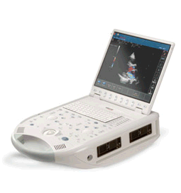 From ESAOTE S.p.A.;
'The MyLab™30CV ultrasound system is an evolutionary step in ultrasound technology. Weighing less than 20 pounds, it is the first compact ultrasound system to deliver premium console performance. And with mobile, portable or stationary configurations, MyLab30CV can adapt to any clinical environment.'
Device Information and Specification
APPLICATIONS
Abdominal, breast, cardiac, OB/GYN, pediatric, pediatric cardiology, small parts, transcranial, vascular
CONFIGURATION
Portable
Linear: 4-10 MHz, convex: 2-5 MHz, phased: 1.6-10 MHz, micro convex: 5-7.5 MHz, endocavity: 5-7.5 MHz, pencil: 2 + 5 MHz
2-D, M-mode, duplex, triplex, color Doppler, pulsed wave Doppler, tissue velocity mapping (TVM), tissue enhancement imaging (TEI™), contrast harmonic imaging, stress echo, tissue velocity mapping for LV motion analysis (TVM), contrast tuned imaging for contrast media procedures (CnTI™), Qontrast™ for myocardium parameters quantification
STORAGE, CONNECTIVITY, OS
Digital patient archive/management, integrated CD/RW, RJ 45 and USB ports, Windows
H*W*D m (inch.)
0.16 * 0.36 * 0.50 (6.2 x 14 x 19.3)
WEIGHT
Less than 11 kg (20 lbs.)
•  From GE Healthcare.;
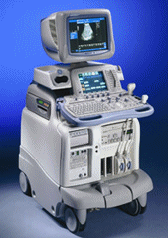
'The System of Choice for General Imaging Imagine a leading-edge ultrasound system so versatile that it can meet the demands of virtually any clinical setting. With the LOGIQ® 9, you'll have a high-performance system capable of multi-dimensional imaging for a full range of clinical applications - from abdominal to breast to vascular imaging. And an ergonomic design that improves scanning comfort and clinical work flow. Now, imagine what LOGIQ® 9 could do for you and your patients.'
Device Information and Specification
APPLICATIONS
Abdominal, cardiac, breast, intraoperative, musculoskeletal, neonatal, OB/GYN, orthopedic, pediatric, small parts, transcranial, urologic, vascular
CONFIGURATION
17' high resolution non-interlaced flat CRT, 4 active probe ports
B-mode, M-mode, coded harmonic imaging, color flow mode (CFM), power Doppler imaging (PDI), PW-HPRF, CW Doppler, color Doppler, pulsed wave Doppler, tissue harmonic imaging
IMAGING OPTIONS
CrossXBeam spatial compounding, coded ultrasound acquisition), speckle reduction imaging (SRI), TruScan technology store raw data, real-time 4D ultrasound, Tru 3D ultrasound
STORAGE, CONNECTIVITY, OS
Patient and image archive, HDD, DICOM 3.0, CD/DVD, MOD, PCMCIA, USB, Windows-based
DATA PROCESSING
Digital beamformer with 1024 system processing channel technology
H*W*D m (inch.)
1.62 * 0.61 * 0.99 (64 * 24 * 39)
WEIGHT
202 kg (408 lb.)
POWER CONSUMPTION
less than 2 KVA
Result Pages : | Share This Page Look Ups |
Medical-Ultrasound-Imaging.com
former US-TIP.com
Member of SoftWays' Medical Imaging Group - MR-TIP • Radiology TIP • Medical-Ultrasound-Imaging
Copyright © 2008 - 2024 SoftWays. All rights reserved.
Terms of Use | Privacy Policy | Advertise With Us
former US-TIP.com
Member of SoftWays' Medical Imaging Group - MR-TIP • Radiology TIP • Medical-Ultrasound-Imaging
Copyright © 2008 - 2024 SoftWays. All rights reserved.
Terms of Use | Privacy Policy | Advertise With Us
[last update: 2023-11-06 01:42:00]